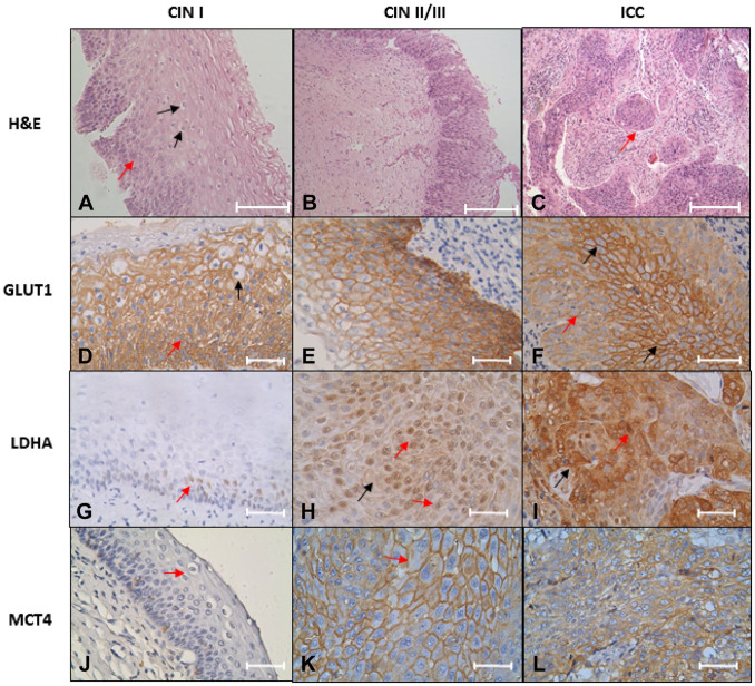
Figure 1.
GLUT1, LDHA, and MCT4 expression in CIN/ICC samples. (A–C) H&E staining. (A) CIN I, dysplastic cell in one third epithelium (red arrow), and some koilocyte cells (black arrow) pathognomonic of HPV infection; (B) CIN II/III, dysplastic cells in all epithelium; (C) invasive non-keratinizing large cell squamous cell carcinoma, tumor nests, are observed (arrow red). Scale bar = 200 µm. (D–F) GLUT1 immunostaining; (D) two third epithelium expression and dysplastic cell (red arrow) and koilocytes (black arrow) with HPV-16, HPV-18,HPV-45, and HPV-6 infection; (E) expression profile in HPV-16-positive infection; (F) expression profile in pleomorphic cells with HPV-16 and HPV-52 infection; a diffuse expression is observed (red arrow). The expression on the cell membrane is present in the expression focus (black arrow). (G–I) LDHA expression; (G) a nuclear expression in the stratum basal cells (red arrow) in HPV-31 and HPV-52 infection samples; (H) nuclear expression (red arrow) and cytoplasm (black arrow) in HPV-39, HPV-51, and HPV-11 infections; (I) nuclear expression (red arrow) and cytoplasm (black arrow) in invasive squamous cell carcinoma, HPV-16 and HPV-39 positive. (J–L) MCT4 immunostaining; (J) CIN I with HPV-16 infection, and koilocyte (red arrow) was observed in the upper layer of epithelium; (K) cytoplasmic membrane of epithelium with multiple infections (HPV-31, HPV-59, and HPV-11) shows a high expression and prominent nucleolus (red arrow). (L) ICC HPV-45 positive indicates many nucleoli. Novolink Polymer. Scale bar = 200 µm for images A–C; scale bar = 50 µm for images D–L. Abbreviations: GLUT1, glucose transporter I; LDHA, lactate dehydrogenase A; MCT4, monocarboxylate transporter type 4; CIN, cervical intraepithelial neoplasia; ICC, invasive cervical carcinoma; HPV, human papillomavirus.

