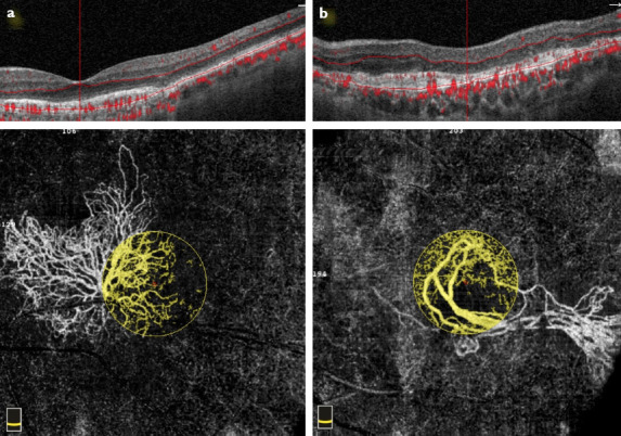
Figure 1.

Optical coherence tomography angiography scan with corresponding OCT B-scan taken at the level of the outer retina to show the morphology of the CNV. (a) Pattern I lesion, the entire extension of a sea-fan Type I CNV is shown: Large mature vessels, branching into tinny capillaries toward the periphery, anastomoses, loops, and peripheral arcades are visible (yellow circle). (b) Pattern II lesion, dead tree appearance Type I CNV is shown: long filamentous linear vessels, branching into rare large mature vessels, with rare or absent anastomoses (yellow circle).
