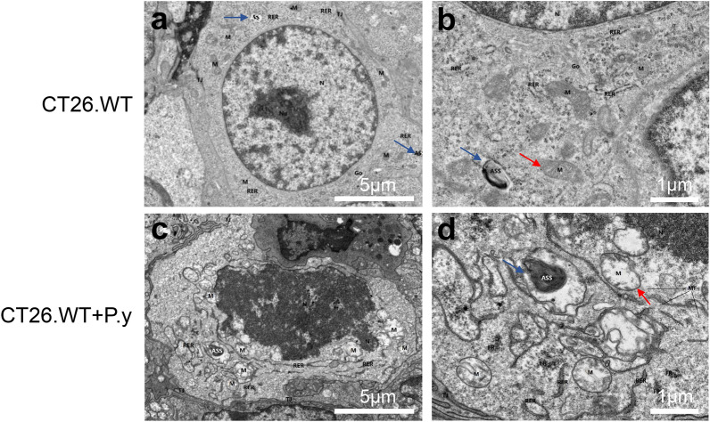
Fig. 5.
Changes in tumor cell ultrastructure after Plasmodium infection. The ultrastructural changes in tumor cells in the two groups were observed by TEM. The damaged cell nuclei and mitochondria were observed in the CT26.WT + P.y group. The mitochondria (M, red arrow) were swollen and the cristae disappeared and vacuolated, with chromatin condensation and nuclear disintegration (N: nucleus). The number of autolysosomes (ASS, blue arrow) in the CT26.WT + P.y group was more than that in the CT26.WT group. a, c The magnification is ×2000, scale bar = 5 µm. b, d The magnification is ×6000, scale bar = 1 µm

