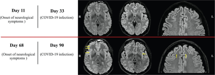
Figure 1.
The trend in neuroradiological detection of white matter changes in our patient is summarised. FLAIR images from the second (top row) and the third (bottom row) MRI examinations. Several new subcortical white matter changes were found and are highlighted with yellow arrows. The discrepancy was not considered to be explained by the slight difference in image quality. All other sequences in all three MR examinations showed normal findings.

