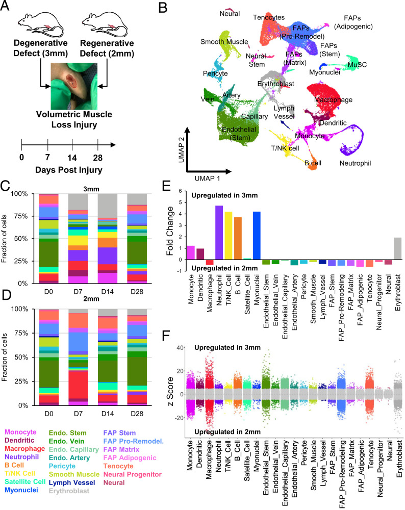
Fig. 2.
scRNA-Seq of regenerative and degenerative muscle defects show exacerbated and persistent inflammation in injuries that do not heal. (A) Schematic of experiment, whereby adult (10 to 12 wk) mice were administered 2-mm or 3-mm biopsy punches to their rectus femoris and humanely euthanized before injury, or 7, 14, or 28 dpi for scRNA-Seq analysis. (B) Dimensional reduction and unsupervised clustering of mononucleated cells isolated from uninjured quadriceps as well as injured quadriceps at 7, 14, and 28 dpi showing 23 different recovered cell types according to marker gene overlays and scCATCH cluster annotation. Two mice were pooled and sequenced for each defect size at each time point, and between 2,337 and 7,500 high-quality libraries were generated for each condition. Quantification of cell abundances at each time point sequenced following (C) 3-mm and (D) 2-mm VML defects shows increased and persistent inflammation among 3-mm defects. (E) Fold changes in cell abundance following 3-mm defects in comparison to 2-mm defects merged across all time points shows nearly fivefold increase in neutrophils, B cells, and T and NK cells. (F) Differential gene expression among each cell type merged across time points and normalized to 2-mm defects. Gray region indicates adjusted P value less than 0.05. Z-scores and P values were calculated for each gene using MAST.

