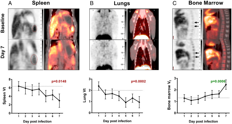
Fig. 2.
DPA-714 binding in EBOV infection. The macaques underwent longitudinal DPA-714 PET imaging at baseline and at the indicated time points postinoculation with EBOV. The representative PET images (Upper) show the changes in DPA-714 binding at baseline and day 7 postinoculation in the (A) spleen, (B) lungs, and (C) bone marrow. The red ovals indicate the splenic region in A. The time course depicting the changes in TSPO binding (Vt) in the organs over time is shown below each image (least-square means adjusted for baseline with 95% confidence intervals). The dotted line represents the average baseline Vt. There is a progressive decrease in TSPO binding in both the spleen and the lungs over time, while there is increased binding seen in the bone marrow. Statistical analysis was performed using the mixed-effect linear regression model and the changes in Vt were found to be correlated with the duration of infection.

