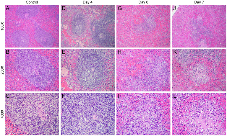
Fig. 4.
H&E spleen (mid disease and terminal). Histopathologic findings for the spleen are consistent with EBOV disease in the nonhuman primate (D–L). Negative controls (A–C, 100×, 200×, and 400×, respectively) show relatively normal splenic architecture. (D–F) 100×, 200×, and 400×, respectively, at day 4 postexposure show mild lymphoid depletion with occasional evidence of lymphocytolysis within germinal centers (white pulp). (G–I) 100×, 200×, and 400×, respectively, at day 6 postexposure show significant lymphoid depletion, with areas of lymphoid degeneration and necrosis within germinal centers, and fibrin with cellular debris in the red pulp. Similarly, in J–L 100×, 200×, and 400×, respectively, at day 7 postexposure, show lymphoid depletion, germinal center necrosis, with hemorrhage, fibrin, and cellular debris in the red pulp. (Scale bars: 100× bar is 100 μ, 200× is 50 μ, and 400× is 20 μ.)

