Fig. 1.
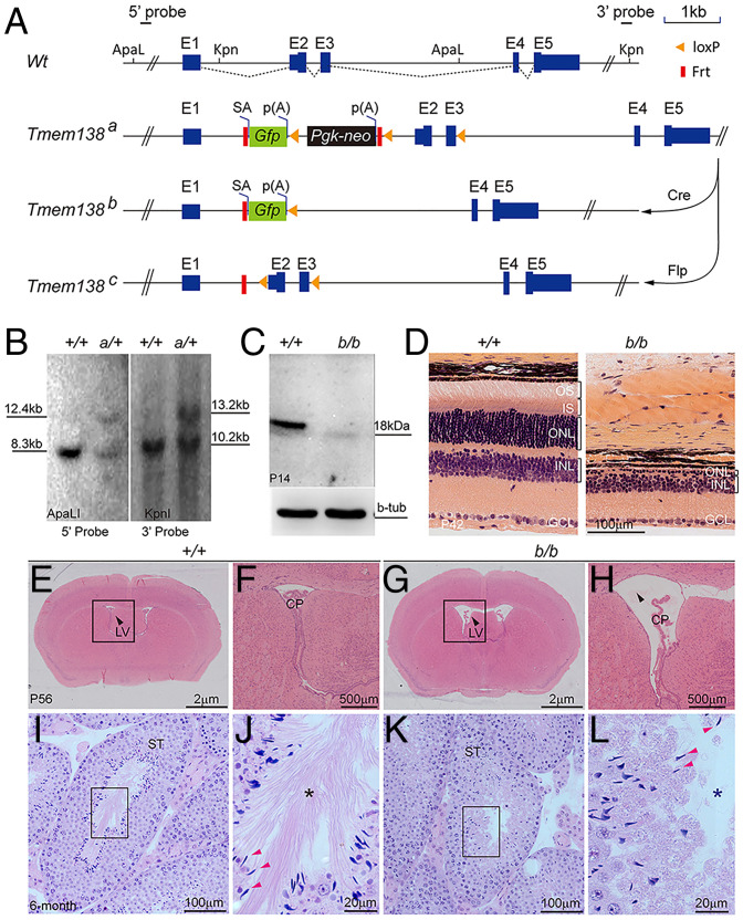
Targeted inactivation of Tmem138 leads to enlarged brain ventricles, azoospermia, and retinal degeneration. (A) Tmem138-targeting strategy. The Tmem138 gene has five exons, with protein coding starting at exon 2 (E2). The restriction enzyme sites ApaLI and Kpn1 were used for Southern blotting analysis in B with 5′ and 3′ probes, respectively; SA, splicing acceptor; p(A), polyadenylation signal; Gfp, green fluorescent protein; Pgk-neo, phosphoglycerate kinase promoter–driven neomycin gene; Flp, flippase; Frt, flippase recognition target. (B) Southern analysis of Tmem138 gene-trap allele (Tmem138a, a/+). The expected fragment sizes on Southern blot are 5′ probe (ApaLI), 8.30 kb (Wt: +/+) and 12.41 kb (a/+), and 3′ probe (KpnI), 10.26 kb (+/+) and 13.28 kb (a/+), respectively. (C) Western blotting of P14 retinal extracts using an anti-Tmem138 antibody; b-tub, β-tubulin. (D) The homozygous Tmem138b/b (b/b)-null mutant manifested severe retinal degeneration at P42; GCL, ganglion cell layer. (E and F) A coronal brain section from a wild-type mouse stained with hematoxylin and eosin at P56 (E). The arrow points to the lateral ventricle (LV). The boxed area is magnified in F; CP; choroid plexus. (G and H) Enlarged brain ventricles of the b/b mice (G). The boxed area is magnified in H. (I–L) Testis sections from 6-mo–old wild-type (I) and null mutant (K) mice, respectively. The boxed areas show the lumen of seminiferous tubes (ST) and are magnified in J and L, respectively. Red arrows point to sperm heads; asterisks indicate sperm tails, which are well developed in the +/+ mice but are absent from the b/b mice.

