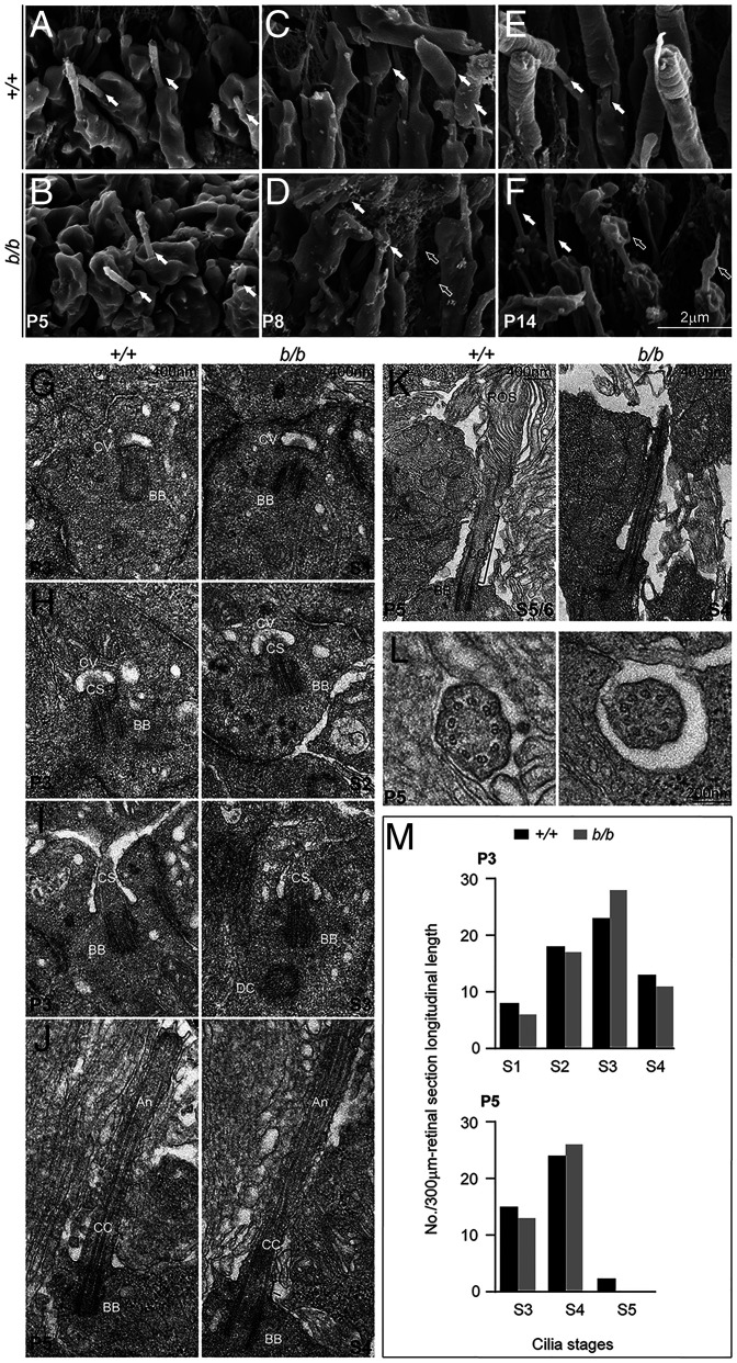
Fig. 6.
Photoreceptor ciliogenesis revealed by SEM and TEM. (A and B) SEM images of wild-type (A) and mutant (B) photoreceptor cilia (arrows) at P5. (C and D) P8 photoreceptors with arrows pointing to the OS in C and membrane material in D. Open arrows point to the membrane vesicles embedded in the interphotoreceptor matrices. (E and F) P14 photoreceptors. Arrows point to the photoreceptor CC; open arrows point to the membranous materials on top of the mutant cilia. (G–J) P3 sections. (G) Stage I cilium (S1), ciliary vesicles docked on the distal end of the mother centrioles. (H) S2 cilium, ciliary shafts elongated within the ciliary vesicles. (I) S3 cilium, ciliary vesicle membrane fused with that of the IS at the apical surface. (J) S4 cilium, cilium grew to assemble axoneme. (K and L) P5 retinal sections. (K) S5/S6, rudimental OS emerged above the cilia in wild-type, but not mutant, photoreceptors; rOS, rudimentary OS. (L) Sections across the CC showed nine doublet microtubules in both wild-type and mutant photoreceptor cells. (M) Quantification of cilia numbers at each stage of P3 and P5. Cilia between RPE (retinal pigment epithelium) and the ONL at each stage of photoreceptor ciliogenesis were counted from three retinal sections/retina, each longitudinally spanning over a 100–μm-length in midperipheral retinal areas. The numbers of cilia from three sections (3 × 100 μm = 300 μm total span) for each stage were used for plotting the graphs. One retina for each genotype was counted. Brackets indicate CC; CV, ciliary vesicle; CS, ciliary shaft. Note that the axonemal, CC, and ciliary shaft are empirically defined according to their relative positions.

