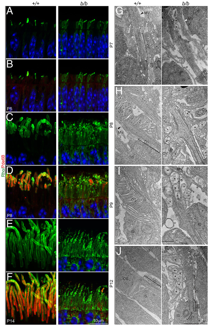
Fig. 7.
Failure of photoreceptor OS formation. (A–F) Photoreceptor OSs were labeled with antibodies to rhodopsin (Rho) and Pde6B. (A and B) Mislocalization of rhodopsin was detected as early as P5 in the mutant retina. Note that Pde6B was only weakly expressed at P5. (C–F) At P8 and P14, rhodopsin and Pde6B were strongly expressed and transported to the OS in the wild-type photoreceptors but were mislocalized throughout the mutant photoreceptor cell body. Note rhodopsin-positive puncta in the presumptive OS and IS regions and the absence of intact OS in the mutant retina. (G–J) TEM observations of photoreceptor OS development; MV, microvilli (arrowheads). Black asterisks indicate aberrant membrane-bound vacuoles; white asterisks indicate vesicles within the ciliary shaft. The black grains in H to J are from nonspecific staining due to sample processing.

