Fig. 10.
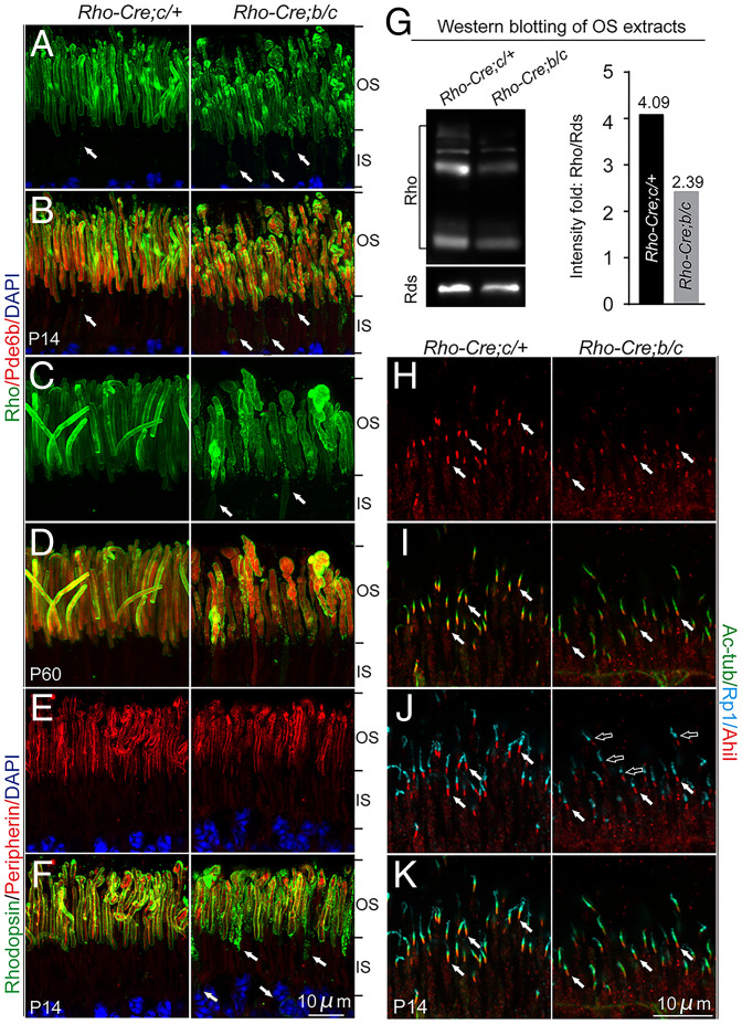
Photoreceptors of Tmem138b/c conditional knockout mice. (A and B) P14 photoreceptors. Disorganized OSs and mislocalization of rhodopsin to the IS were revealed by rhodopsin (green) and Pde6b (red) staining. (C and D) Two–mo-old photoreceptors. The mutant photoreceptors appeared swollen. Arrows in A to C point to mislocalized rhodopsin. (E and F) Colabeling of peripherin/Rds (red) and rhodopsin (green) at P14. (G) Western blotting of rhodopsin and Rds using P21 photoreceptor OS extracts (Materials and Methods). Right, quantification of the rhodopsin/Rds signal intensity fold. (H–K) P14 photoreceptors. Shortened and diminished Ahi1 (red) and Rp1 (turquoise) staining was observed. White arrows point to the Ahi1 staining. Open arrows point to the gap between Ahi1 and Rp1 in the mutants.

