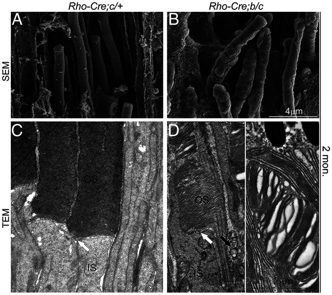
Fig. 11.
SEM and TEM of the photoreceptors of Tmem138b/c conditional knockout mice. (A and B) SEM images of wild-type (A) and mutant (B) photoreceptor OSs. (C and D) TEM images of wild-type (C) and mutant (D) photoreceptor OSs. White arrows point to the base (nascent disc) of the OS. The black arrow in D indicates the mutant photoreceptor extracellular vesicles. The bracket indicates variable sizes of intracellular vesicles within the OS. Retinal sections were prepared from 2–mo-old mice.

