Fig. 7.
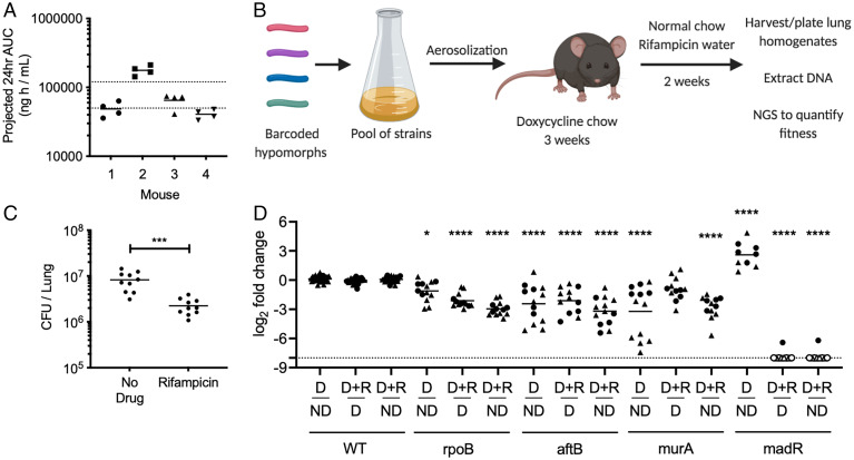
Essential bacterial functions that alter drug efficacy in vivo. (A) RIF plasma concentrations were measured in mice over a 12-h period, 24 h post-RIF administration (0.1 g/L). Results shown as area under the concentration (AUC) time profile. Dotted lines indicate RIF plasma range observed during clinical TB therapy (58). (B) C57BL/6J mice were infected through the aerosol route with a pooled culture of individual hypomorph mutants and barcoded WT strains. Mice were fed doxycycline chow starting 3 d before infection to 3 wk postinfection. Mice were subsequently switched to normal chow and water with or without RIF (0.1 g/L) for 2 additional weeks. Lungs were harvested, homogenized, and plated for Mtb outgrowth. Upon DNA extraction of grown colonies, chromosomal barcodes were PCR amplified and pooled for Illumina NGS. Barcode abundances of individual mutants were normalized to WT strains and analyzed to quantify changes in fitness during RIF treatment. (C) RIF-treated mice show significant decrease in Mtb CFU in lungs compared to untreated controls. Each dot represents a single mouse. Significance was calculated using unpaired t test, ***P < 0.001. (D) Change in fitness of individual mutants was determined by comparing normalized counts from nondepleted (ND), depleted (D), as well as depleted and RIF-treated (D+R) conditions. Depletion of RpoB, AftB, and MurA display significant decrease in fitness, shown as log2 fold change, compared to nondepleted controls. Depletion of RpoB, AftB, and MadR show significant decrease in fitness during RIF treatment compared to depleted but untreated controls. Results shown as two independent experiments (circle and triangle) with each dot representing a single mouse. Empty dot indicates that the relative abundance was below the limit of detection (dotted line). Significance was calculated using Sidak’s multiple comparison test, *P < 0.05, ****P < 0.0001.

