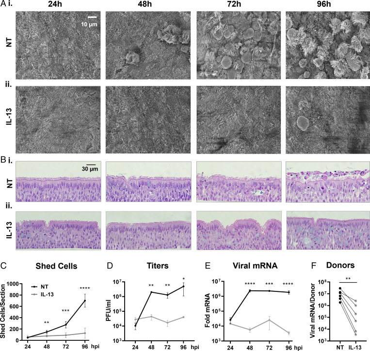
Fig. 4.
Effects of IL-13 treatment on viral replication and cell shedding. Three days prior to infection, HAE cultures were divided into two groups, the NT/PBS group (NT) and the IL-13–treated group (IL-13) to which 1 ng/mL of IL-13 was added via the basolateral side. On day 0, cells were infected with SARS-CoV-2 (MOI = 0.5) and processed at 24, 48, 72, and 96 hpi for histology, viral titers, and mRNA extraction. (A) Representative SEM en face views of infected HAE cells at 24, 48, 72, and 96 hpi for NT (i) and IL-13–treated (ii) cells. Viral shedding, cell swelling, and detachment was observed at 24 hpi to 48 hpi in the NT group, while limited epithelial damage was noticed up to 96 hpi in the IL-13 group. (B) Representative H&E images of infected HAE cells at 24, 48, 72, and 96 hpi for NT (i) and IL-13–treated (ii) cells. (C) Graph showing the number of shed cells per histological section for NT (black) and IL-13 (gray) groups; n = 4 sections per group. (D) Progression of viral titers in PFU per milliliter over time in NT and IL-13–treated cells; n = 3 inserts per time point per group. (E) Relative change in the nucleocapsid gene mRNA expression for both groups. Fold change shown as 2^-ΔΔCt; n = 3 cultures per time point per group. (F) Paired graph for the change in nucleocapsid mRNA expression at 96 hpi in response to IL-13 treatment for six donors; n = 3 to 6 inserts per group per donor. *P < 0.05, **P < 0.01, ***P < 0.001, ****P < 0.0001.

