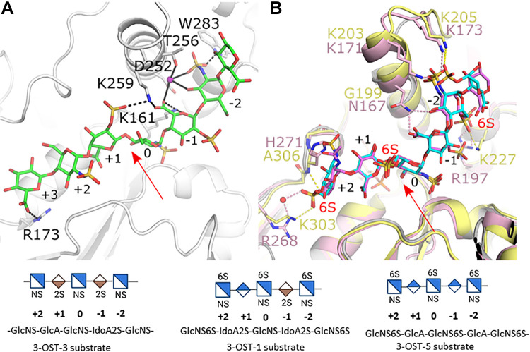
Figure 3.
Differences in HS binding between the 3-O-sulfotransferase (3-OST) isoforms -1, -3, and -5. A: Crystal structure of the catalytic domain of 3-OST-3 binding oligosaccharide substrate (protein gray, HS green; pdbcode 6XL8). B: superposition of crystal structures of 3-OST-1 (pink, substrate dark pink; PDBcode 3UAN) and -5 (light yellow, substrate cyan; pdbcode 7SCE). Residues discussed in text are displayed in stick. Red arrows denote the acceptor 3-OH on glucosamine 0. Pink, black, and yellow dashed lines represent interactions between HS and 3-OST-1, -3, and -5, respectively. Solid black lines represent interactions in 3-OST-3 with the sodium ion. Shorthand representation of the substrates in the active site are shown at the bottom of the figure. HS, heparan sulfate.

