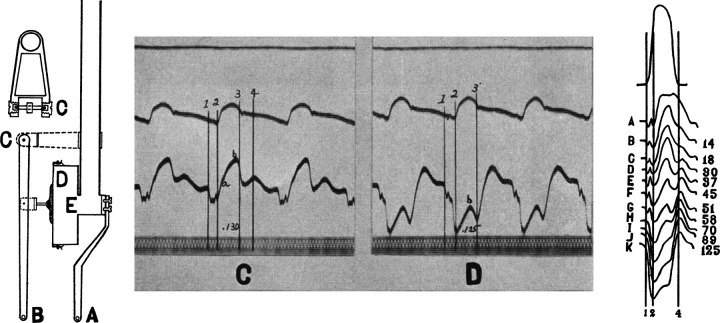
Figure 8.
Tenant and Wiggers’ (81) original description of the effects of brief ischemia on cardiac contraction. Left: innovative optical myograph used to assess cardiac contraction (strain) during an acute occlusion of the left anterior descending coronary artery. Middle: original recordings of aortic pressure and dynamic myocardial strain throughout that cardiac cycle on a beat-to-beat basis during a coronary occlusion. Vertical bars indicate 1) end-diastole, 2) onset of ejection, 3) end-systole or aortic valve closure, and 4) onset of diastole and mitral valve opening. Systolic shortening at rest (C) transitions to lengthening or dyskinesis during brief ischemia (D). Right: left ventricular pressure below which are strain measurements demonstrating the rapid and progressive reductions in systolic shortening on a beat-to-beat basis during 125 s of ischemia. Dyskinesis and systolic lengthening consistently developed by 1 min of ischemia. Adapted from Tennant and Wiggers (81) and reproduced with permission of the American Physiological Society.

