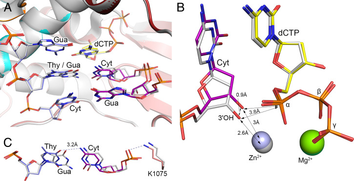
Fig. 1.
Close-up view of the human Polα active site in the aligned ternary complexes containing a matched and mismatched template:primer. (A) Close-up view of the postinsertion and adjacent sites in the aligned ternary complexes. (B) The 3′-OH of the mismatched primer is shifted by 0.9 Å toward the template. The primer 3′-OH was modeled using the Builder tool in the PyMOL Molecular Graphics System. (C) Aligned G-C base pair and T-C mispair at the postinsertion site of PolαCD. The hydrogen bonds in the complex with a T-C mismatch are indicated by light-blue dashed lines. In the complex with a mismatched template:primer, the carbons of dCTP, DNA template, and RNA primer are colored yellow, marine, and purple, respectively. The subdomains of PolαCD—N-terminal, fingers, and palm—are colored orange, cyan, and salmon, respectively. In the complex containing a correct template:primer (PDB code 4qcl), all molecules are colored gray with 10% transparency. Magnesium and zinc ions in the structure with a matched duplex are colored gray and dark green, respectively. The complexes are aligned with an rmsd of 0.075 Å for 850 Cα atoms.

