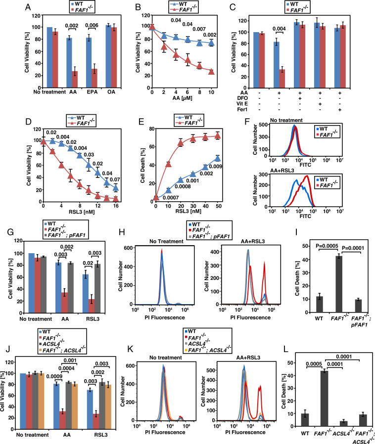
Fig. 1.
FAF1 deficiency sensitizes cells to ferroptosis. (A–D, G, and J) Viability of SV589-derived cells treated with indicated FAs (10 µM, indicated concentration in B), deferoxamine (DFO, 10 µM), vitamin E (Vit E, 2 µM), ferrostatin 1 (Fer1, 0.1 µM), RSL3 (D, indicated concentration; G and J, 10 nM) for 24 h was measured as described in Materials and Methods, with the value of the untreated WT cells set at 100%. (E) The percentage of dying cells determined through flow cytometry of the cells treated with 10 µM AA and the indicated amount of RSL3 for 12 h followed by propidium iodide (PI) staining. (F) BODIPY 581/591 C11 flow cytometry of the green fluorescence of indicated cells incubated with or without 10 µM AA for 4 h followed by cotreatment with 50 nM RSL3 for 2 h. (H and K) Flow cytometry of cells treated with 10 µM AA and 10 nM RSL3 for 12 h followed by PI staining. (I and L) The percentage of dying cells in experiments shown in H and K, respectively. (A–E, G, I, J, and L) Results are reported as mean ± SEM from three independent experiments. Statistical significance was calculated by unpaired, two-tailed t test.

