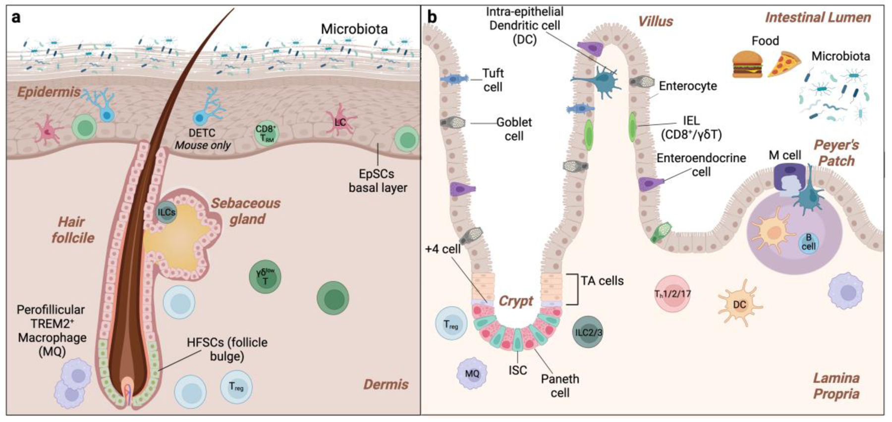
Figure 1. Epithelial and Immunological features of the Skin and Gut.

a. Basal epidermal stem and progenitor cells (EpSCs) EpSCs differentiate upward to sustain the interfollicular epidermis and hair follicle stem cells (HFSCs) residing in the bulge fuel follicle cycling. The immune niche of the epidermis includes basal Langerhans cells (LCs), CD8+ resident memory T cells (TRM), innate lymphoid cells (ILCs), and, in murine skin, suprabasal Dendritic Epidermal γδ T cells (DETCs). Regulatory T cells (Tregs) and macrophages are located in the perifollicular dermis. b. The intestinal epithelium is organized in autonomous crypt-villus structures. Intestinal stem cells (ISCs) located at the base of the crypt maintains proliferate and differentiate upward to maintain each unit. +4 cells are a reserve SC population that can substitute for ISCs upon depletion. Intestinal progenitors in the transit-amplifying (TA) region further differentiate into the six lineages of absorptive (M-cells and enterocytes) and secretory (goblet cells, enteroendocrine cells, tuft cells, and Paneth cells) intestinal cells. The intestinal immune niche includes intraepithelial lymphocytes (IELs) and DC (intraepithelial DCs), Treg, Th, γδ T cells, B cells, DCs, and macrophages. Peyer’s patch are lymphoid aggregates that are play vital role in immune surveillance. M cells sample luminal antigens and live bacteria and transcytose across the epithelial barrier to DCs.
