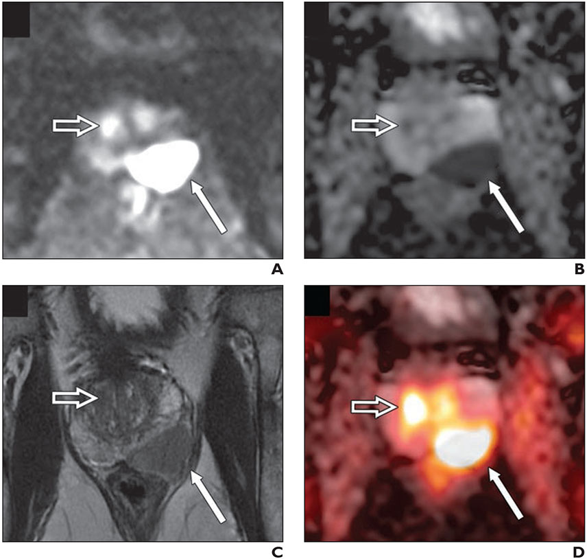
Fig. 2—
77-year-old man with biopsy-proven Gleason score 4 + 5 prostate cancer in left base and serum PSA level of 8.4 ng/mL with negative conventional staging (no evidence of metastatic disease).
A–C, DWI (b value, 2000 s/mm2) (A), apparent diffusion coefficient map (B), and small FOV T2-weighted MRI (C) images show large PI-RADS 5 lesion in left base with T2-weighted hypointensity and marked restricted diffusion (thin arrow). Open arrow shows uptake in transition zone.
D, Fused 18F-fluciclovine PET/MRI shows high focal activity within lesion (thin arrow). Second focus of uptake (open arrow) in transition zone with no anatomic lesion on T2-weighted images and mild corresponding restricted diffusion corresponds to benign prostatic hyperplasia nodule.

