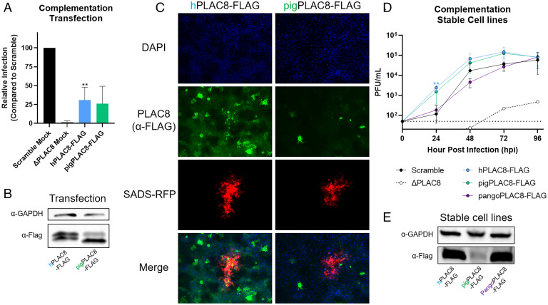
Fig. 5.
Complementation with ectopic or stable PLAC8 expression rescues SADS-CoV infection. (A) PLAC8 KO cells were transiently transfected with human and pigPLAC8-FLAG (codon optimized). Subsequently, transfected cells were infected with SADS-CoV-nLuc (MOI: 0.015) for 48 hpi. RLUs from the infected cells were used as surrogates for viral infection. (B) Western blot images of hPLAC8 and pigPLAC8-FLAG expression on Huh7.5 cells after transient transfection using anti-FLAG antibody. (C) Representative immunofluorescence images of hPLAC8- and pigPLAC8-FLAG–transfected cells infected with SADS-CoV-RFP and stained with anti-FLAG antibody (green), SADS-CoV-RFP (red), and DAPI (blue). (D) SADS-CoV-RFP growth kinetics on Huh7.5 ΔPLAC8 cells stably expressing hPLAC8, codon-optimized pigPLAC8, and pangoPLAC8-FLAG. (E) Western blot images of hPLAC8, pigPLAC8, and pangoPLAC8-FLAG expression in stable cell lines using anti-FLAG antibody. Error bars represent ± 1 SD, *P < 0.05, **P < 0.01, ***P < 0.001 by Student's t test.

