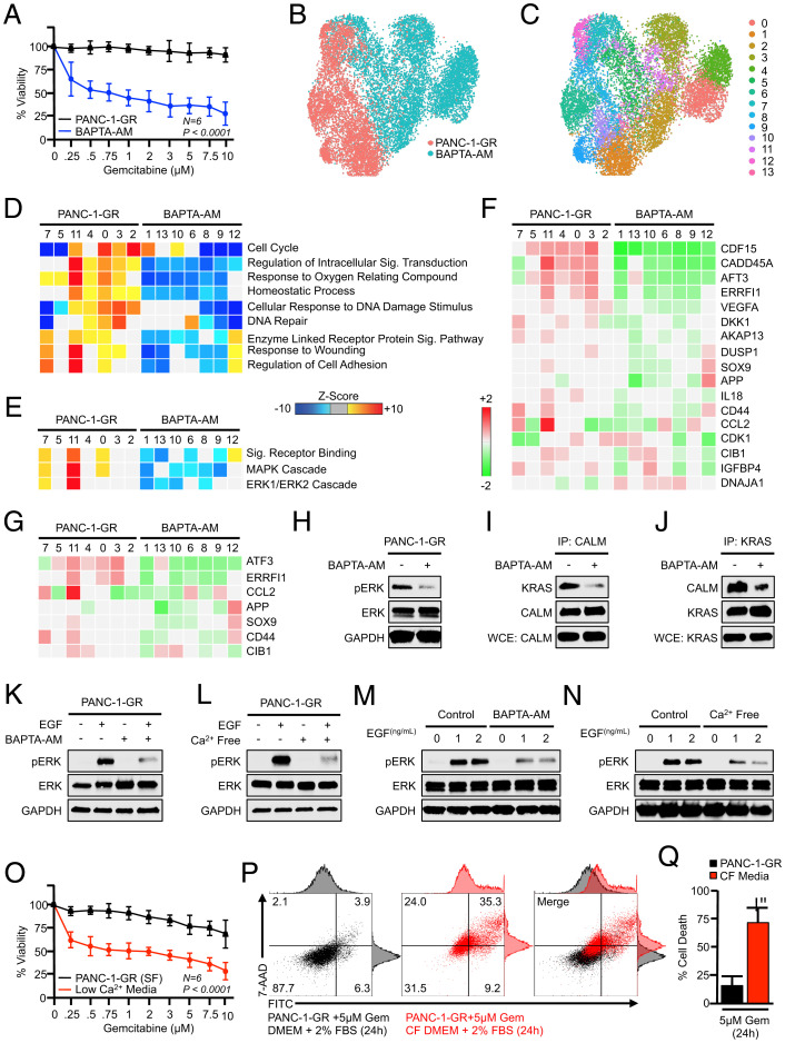
Fig. 4.
Calcium depletion disrupts ERK activation and restores gemcitabine sensitivity in vitro. (A) PANC-1-GR cells were incubated with a fixed concentration of either a DMSO control (1:1,000) or the calcium chelator BAPTA-AM (10 μM). After 2 h, cells were challenged with increasing concentrations of gemcitabine, and cell viability was evaluated after 48 h by MTT assay. Error bars represent mean ± SEM. (B–E) PANC-1-GR cells were treated with either a DMSO vehicle or BAPTA-AM (10 μM) for 24 h. Cells were then collected and evaluated by single-cell RNA sequencing. Cell populations were visualized via UMAP scatterplot, transcriptionally distinct clusters were identified, and each was subjected to enrichment analysis for cell processes and signaling pathways identified previously, with BAPTA-AM–treated clusters showing pronounced down-regulation of MAPK and ERK signaling. (F and G) Individual genes in the ERK and MAPK gene sets, respectively. (H) PANC-1-GR cells were treated with either a DMSO vehicle or BAPTA-AM (10 μM) for 24 h, after which ERK activation was assayed by Western blot. (I and J) PANC-1-GR cells were treated similarly, and the interaction between KRAS and CALM was evaluated by immunoprecipitation. (K and L) PANC-1-GR cells were incubated with either a DMSO vehicle or BAPTA-AM in serum-free media for 24 h. Cells were then stimulated with 0.5 ng/mL recombinant EGF, and ERK activation was evaluated by Western blot after 10 min. The experiment was then repeated using cells grown in either control serum-free media or calcium (Ca2+)-free, serum-free media. (M and N) This experiment was repeated using 0, 1, or 2 ng/mL EGF. (O) PANC-1-GR cells were cultured overnight in either unmodified serum control, serum-free media, or Ca2+-free, serum-free media. At this time, cells were challenged with increasing concentrations of gemcitabine, and cell viability was evaluated after 24 h by MTT assay. Error bars represent mean ± SEM. (P and Q) PANC-1-GR cells were grown in either control media supplemented with 2% FBS or Ca2+-free (CF) media with 2% FBS (low-Ca2+ media) and cell death was evaluated by Annexin-FITC assay. Error bars represent mean ± SEM (*P < 0.05).

