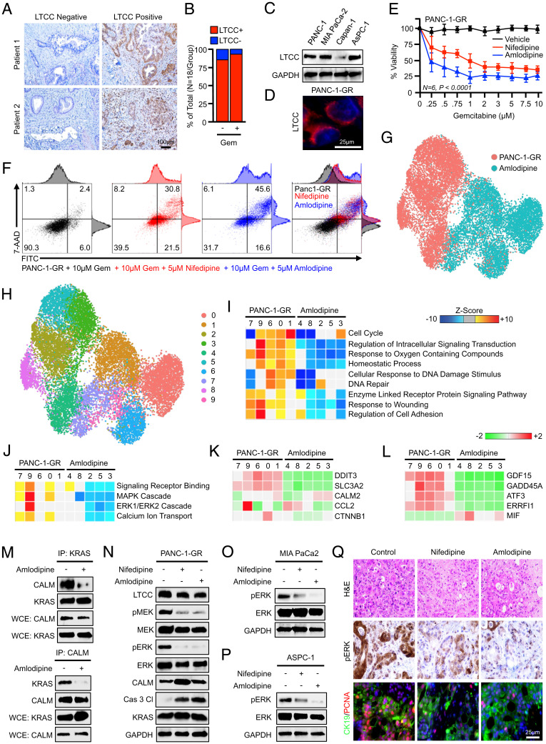
Fig. 5.
Calcium channel blockers impair prosurvival ERK signaling and improve gemcitabine sensitivity in vitro. (A) Excisional biopsies from 36 PDAC patients were sectioned and stained via immunohistochemistry for LTCCs and representative images are shown for each from either chemotherapy-naïve patients (n = 18) or patients who had received neoadjuvant gemcitabine-based chemotherapy (n = 18). (B) The percentage of patients in each group with LTCC-expressing and non–LTCC-expressing tumors. (C) PANC-1, MiaPaCa-2, Capan-1, and ASPC-1 cells were evaluated for LTCC expression by Western blot. (D) PANC-1-GR cells were stained for LTCCs by immunofluorescence, showing strong membrane localization. (E) PANC-1-GR cells were incubated with a fixed concentration of either a DMSO vehicle (1:1,000) or the CCBs nifedipine (5 μM) or amlodipine (5 μM). After 2 h, cells were challenged with increasing concentrations of gemcitabine, and cell viability was evaluated after 48 h by MTT assay. Error bars represent mean ± SEM. (F) PANC-1-GR cells were treated similarly and cell death was evaluated by Annexin-FITC assay. (G–J) PANC-1-GR cells incubated with either a DMSO control vehicle or amlodipine (5 μM) for 24 h. Cells were then collected and evaluated by single-cell RNA sequencing. Cell populations were visualized via UMAP scatterplot, transcriptionally distinct clusters were identified, and each was subjected to enrichment analysis for cell processes identified in Fig. 1, with amlodipine-treated clusters showing significant down-regulation of MAPK and ERK signaling. (K and L) Individual genes in the ERK and MAPK gene sets, respectively. (M and N) PANC-1-GR cells were treated similarly, and the interaction between KRAS and calmodulin was evaluated by immunoprecipitation. Cell lysate was also evaluated by Western blot for ERK pathway activation. (O and P) MiaPaCa-2 and ASPC-1 cells were treated with nifedipine (5 μM) or amlodipine (5 μM), and ERK activation was evaluated by Western blot. (Q) Excisional biopsies from two PDAC patients undergoing survival resection were cored, sectioned at 250-μm intervals, and cultured ex vivo either in a control DMSO vehicle, nifedipine (5 μM), or amlodipine (5 μM). After 72 h, slice cultures were formalin-fixed, paraffin-embedded, and stained either with H&E, via immunohistochemistry for CALM, or dual-stained for CK19 and PCNA.

