Fig. 7.
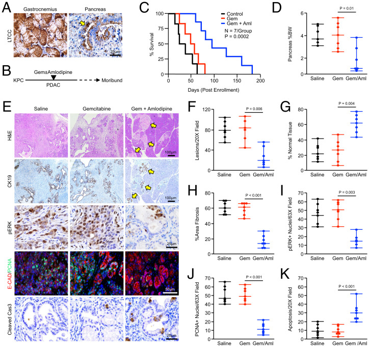
Amlodipine potentiates gemcitabine chemotherapy in KPC mice. (A) Pdx1-Cre × LSL-KrasG12D × LSL-TP53R172H(KPC) mice were generated as a model of advanced PDAC. Pancreas tissues were collected from tumor-bearing mice, sectioned, and stained via immunohistochemistry for LTCCs, with muscle tissue from the gastrocnemius used as a positive control. The arrows indicate LTCC-expressing neoplastic tissues. (B and C) Starting at 15 wk of age, KPC mice were enrolled into one of four treatment groups. Mice were either treated with IP injections of a saline vehicle, 100 mg/kg gemcitabine twice per week, daily injections of 2 mg/kg amlodipine, or gemcitabine and amlodipine. Mice were killed when showing clear signs of health decline such as weight loss or lethargy, and survival is shown via the Kaplan–Meier method. For the amlodipine monotherapy group, see SI Appendix, Fig. S9A. (D) At the study end point, the pancreas gland was weighed and normalized to each animal’s body weight (BW), and results are displayed as individual value plots. For the amlodipine monotherapy group, see SI Appendix, Fig. S9B. (E–K) Pancreas tissues were stained with H&E or via immunohistochemistry for CK19, pERK, E-cadherin and PCNA, or cleaved caspase 3. Tissues were quantified as described and results are displayed as individual value plots. For the amlodipine monotherapy group, see SI Appendix, Fig. S9 C–H.

