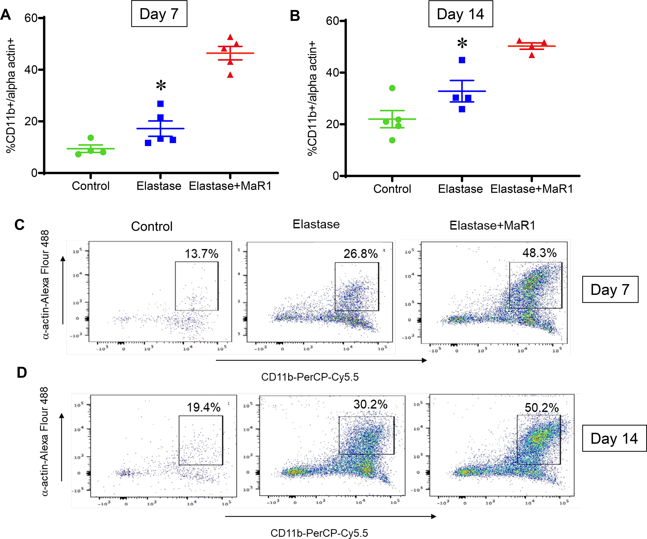
Figure 5.

MaR1 increases efferocytosis of SMCs in aortic tissue of murine AAA. Flow cytometry analysis of murine aortic tissue demonstrated increased levels of co-expression CD11b+SM-αA+ cell population in MaR1 treated mice compared to mice treated with vehicle alone at (A) post-operative day 7 (*p<0.001, n=4–5 per group) and (B) post-operative day 14 (*p=0.01, n=4–5 per group). Representative flow cytometry panels of day 7 (C) and day 14 (D) murine aortic tissue analysis.
