. 2022 May 23;11:e66909. doi: 10.7554/eLife.66909

Figure 1. Dnmt3a conditional knockout (cKO) in cortical pyramidal neurons during mid-gestation impaired working memory, social interest, and acoustic startle responses.

(A) An experimental model of the conditional loss of Dnmt3a in excitatory neurons. P0 and P39, postnatal day 0 and 39. FANS, fluorescence-activated nuclei sorting. (B) RNA-seq confirmation of the deletion of Dnmt3a exon 19 in P39 excitatory neurons. RPKM, reads per kilobase per million. R1/2, replicate 1/2. *, t-test p=0.014. (C) Dnmt3a cKO mice made fewer spontaneous alternations in the Y-maze test of working memory (Wilcoxon test, **, p=0.0079; *, p=0.011; n=15 male control, 15 male cKO, 11 female control, 10 female cKO). (D) Male Dnmt3a cKO mice spent less time interacting with an unfamiliar mouse, indicating reduced social interest (Wilcoxon test; *, p=0.01048; **p=0.006833; n=14 male control, 15 male cKO, 11 female control, 10 female cKO). (E) Male Dnmt3a cKO mice had decreased startle response to a 120 dB acoustic pulse (Wilcoxon test, **, p=0.0019; n.s., not significant; n=14 male control, 15 male cKO, 11 female control, 10 female cKO).

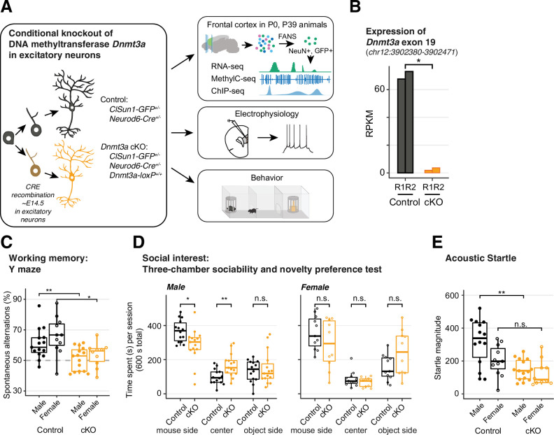
Figure 1.

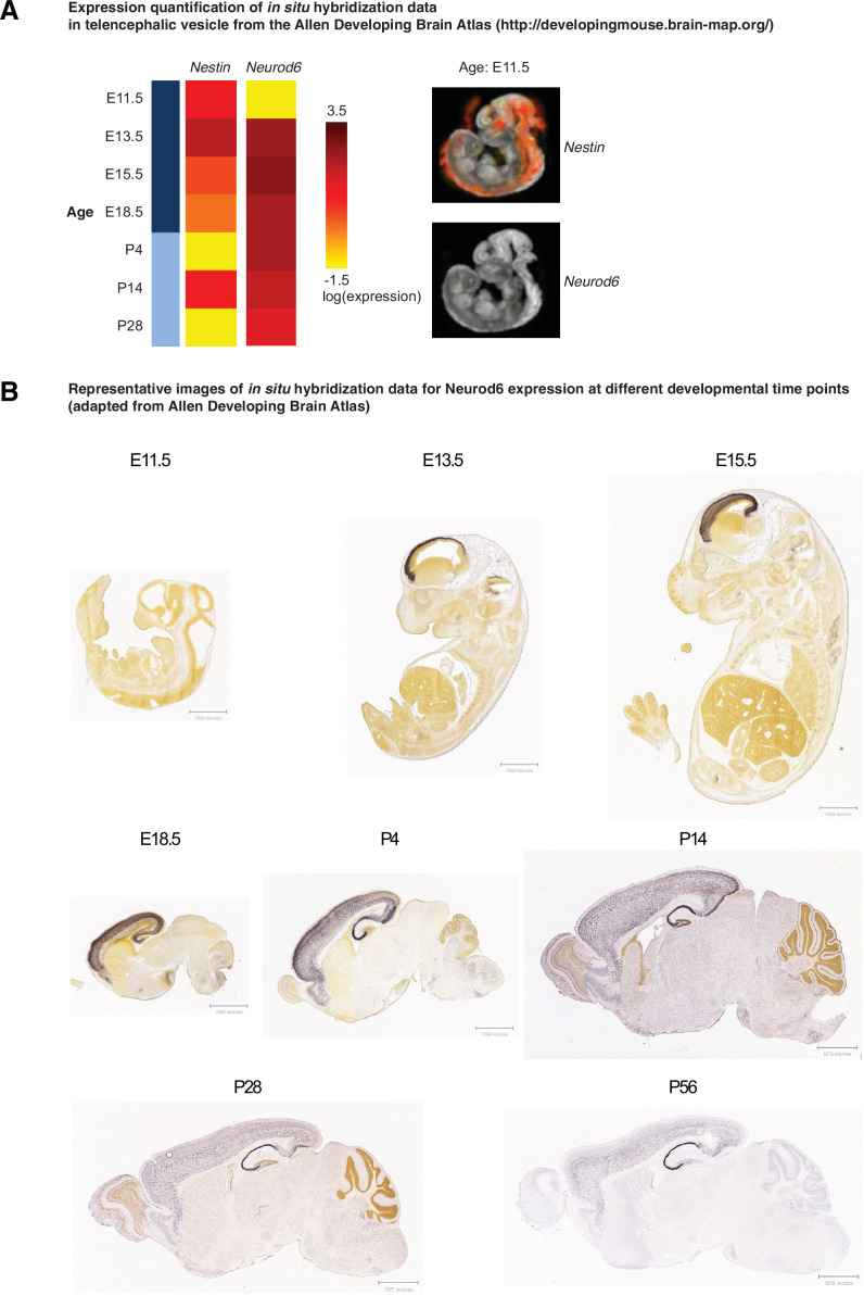
Figure 1—figure supplement 1. Neurod6 starts to express between embryonic day E11 and E13.

Figure 1—figure supplement 1.

(A) Expression quantification of in situ hybridization data of gene Nestin and Neurod6 in telencephalic vesicle from the Allen Developing Brain Atlas (http://developingmouse.brain-map.org/). Left panel, heatmap of the gene expression across ages during development. Right panel, example images of the gene expression in E11.5. Image credit: Allen Institute. E11.5–18.5, embryonic days; P4–28, postnatal days. (B) Expression of Neurod6 at different developmental time points (embryonic days 11.5, 13.5, and 15.5, and postnatal days 4, 14, 28, and 56). Images were taken from the Allen Developing Brain Atlas.
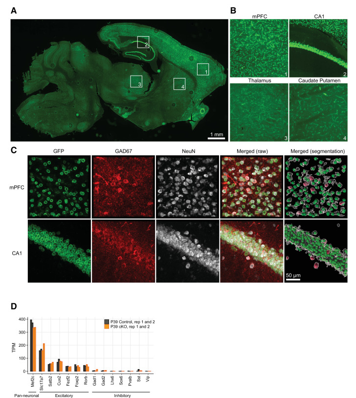
Figure 1—figure supplement 2. Neurod6-dependent Cre recombination occurred only in excitatory neurons.

Figure 1—figure supplement 2.

(A–C) Cell-type-specific expression of nuclear membrane tag (Sun1-sfGFP-myc) in non-inhibitory NEUROD6+ neurons in mouse brain. (A) Overview (10× magnification) of a sagittal section stained with anti-GFP antibody. Note that the Sun1-sfGFP nuclear tag expression is region-specific, as described for the characterization of the NexCre mouse line (Goebbels et al., 2006). The numbered insets in (A) are enlarged and shown in (B), as example regions where the nuclear membrane tag is expressed (mPFC and CA1 hippocampal region) and where it is not (caudate putamen and thalamus). (C) Confocal images (60× magnification, maximum intensity projection) of mouse brain slices (40 µm thick) triple-labeled with antibodies anti-GFP (Sun1-sfGFP-tag, green channel), anti-GAD67 (inhibitory cells marker, red channel), and anti-NeuN (neuronal marker, gray). Note that Sun1-sfGFP-tag is only expressed in neurons that are not inhibitory. (D) Expression of pan-neuronal, excitatory, and inhibitory neuron marker genes in our RNA-seq data. TPM, transcripts per million.
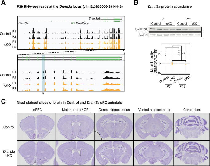
Figure 1—figure supplement 3. Dnmt3a was disrupted on both the mRNA and protein levels in the Dnmt3a conditional knockout (cKO) excitatory neurons.

Figure 1—figure supplement 3.

(A) Genome browser tracks of mRNA-seq data show confirmation of the deletion of Dnmt3a exon 19 in P39 Dnmt3a cKO excitatory neurons. The targeted exon region is highlighted in the light blue shaded box with an asterisk. R1/2, replicate 1/2. (B) The protein product of the Dnmt3a gene is disrupted in the cKO sample. Top panel, Western blot; Bottom panel, quantification of the protein abundance. P5 and P13, postnatal days 5 and 13. **, t-test p=0.0017. (C) Nissl-stained slices show no morphological alterations in the brain of the Dnmt3a cKO animals. mPFC, medial prefrontal cortex; CPu, caudate putamen.
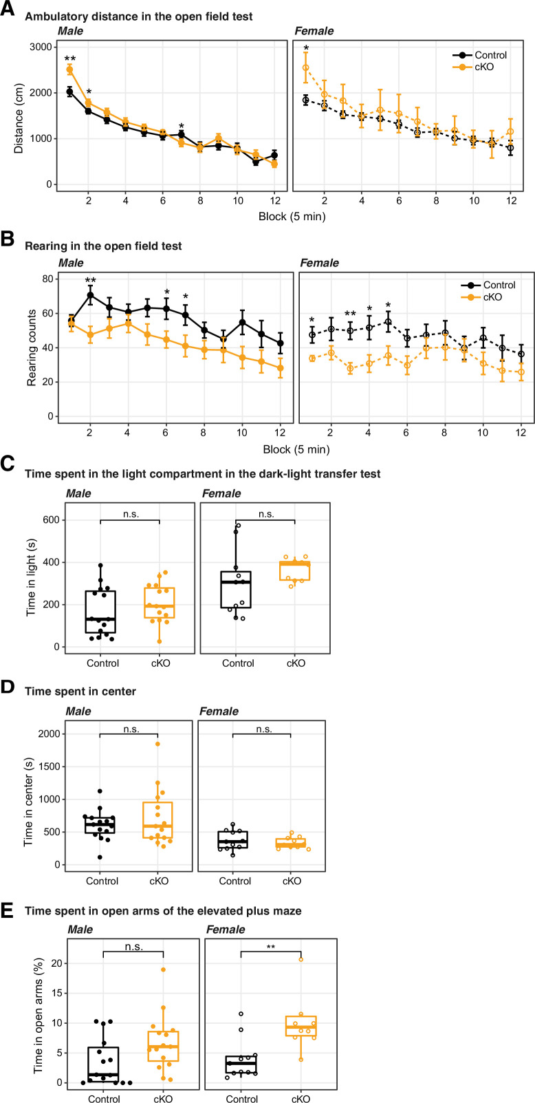
Figure 1—figure supplement 4. The conditional ablation of Dnmt3a in pyramidal neurons did not significantly impair motor activity nor increased anxiety levels.

Figure 1—figure supplement 4.

(A) and (D) Dnmt3a conditional knockout (cKO) mice displayed normal behavior in the open field test, traveling a similar distance (A) as control mice, and also showing a similar degree of center activity (D) (B) The exploratory activity was slightly decreased in Dnmt3a cKO animals, as suggested by an attenuated rearing behavior. (C) The time spent in light in the dark-light transfer test was not significantly affected by the lack of Dnmt3a. (E) The female, but not the male, cohort of Dnmt3a cKO mice spent significantly more time than control mice in the open arms of the elevated plus maze, consistent with lower anxiety levels (Wilcoxon test, **, p=0.0048; n.s., not significant.). In the line plots (A–B), data were presented as mean ± SEM In all boxplots (C–E), the middle horizontal bar represents the median; the lower and upper hinges correspond to the first and third quartiles, and the whisker extends from the hinge to the value no further than 1.5 * IQR from the hinge, where IQR is the interquartile range. The values of individual experiments are represented by dots superimposed on the boxplots. Wilcoxon test significance: *, p<0.05; **, p<0.01; n.s., not significant.
Figure 1—figure supplement 5. Dnmt3a conditional knockout (cKO)-induced impairment of startle response was accompanied by increased prepulse inhibition (PPI), and the cKO did not affect fear memory.

Figure 1—figure supplement 5.

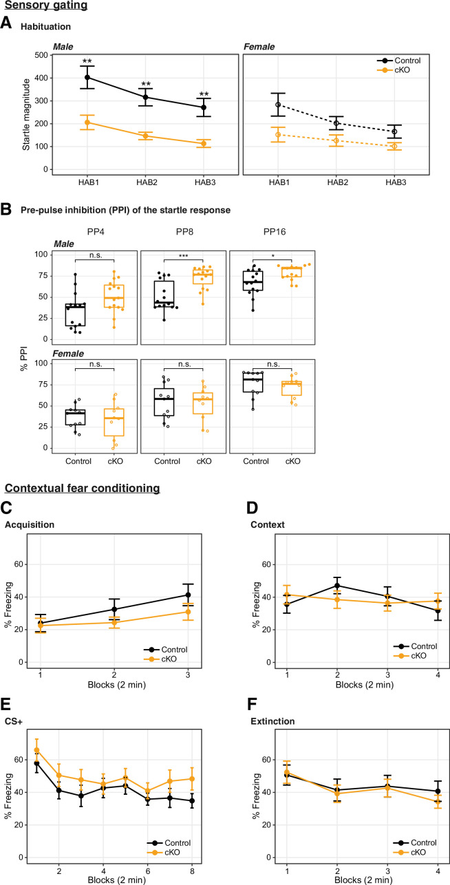
(A) The increased PPI accompanied the impairment in startle responses to a 120 dB tone played at three time points during the recording session (HAB1 – beginning of the session; HAB2 – middle of the session; HAB3 – end of the session) (Wilcoxon test p=0.0027, 0.0019, and 0.0035 in male HAB1, HAB2, HAB3, respectively, and not significant in female). The habituation to the 120 dB auditory tone (i.e. the relative reduction in startle response throughout the experiment) was not significantly different between genotypes. (B) The percentage of PPI at prepulse intensity of 69, 73, and 81 dB (4, 8, and 16 dB above the 65 dB background, respectively) was increased in male, but not female mice (Wilcoxon test, ***, p=0.00076; *, p=0.016; n.s, not significant). (C–F) Fear learning and extinction were tested over 4 consecutive days. N=14–15 per group. (C) Fear acquisition to three tone-shock pairings occurred on day 1; (D) contextual fear in relation to the acquisition context (8 min, Block = 2 min) was measured on day 2; (E) cued fear recall and extinction training occurred on day 3 (Block = 4 tone trials); and (F) extinction recall (Block = 4 tone trials) occurred on day 4. Wilcoxon test reported no significant changes between the Dnmt3a cKO and control. In boxplots (A), the middle horizontal bar represents the median; the lower and upper hinges correspond to the first and third quartiles, and the whisker extends from the hinge to the value no further than 1.5 * IQR from the hinge, where IQR is the interquartile range. The values of individual experiments are represented by dots superimposed on the boxplots. In the line plots (B–F), data were presented as mean ± SEM.