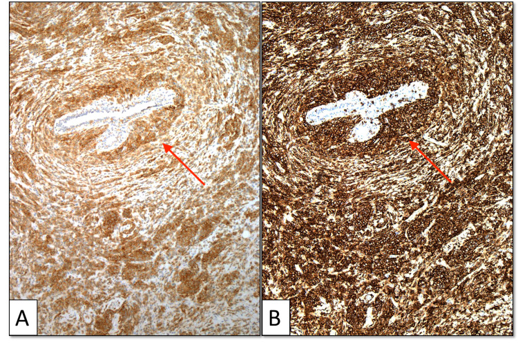
Figure 4. CD33 and CD43 immunohistochemistry of the core biopsy specimen.
CD33 and CD43 immunohistochemistry with original ×100 magnification. There is diffuse positive cytoplasmic staining of myeloid cells for CD33 (A) and CD43 (B). A tumor surrounds and focally infiltrates a benign breast duct (arrows).

