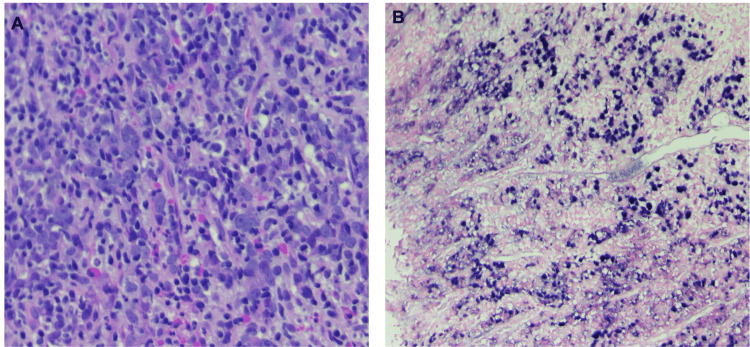
Figure 1. (A) Poorly differentiated large and oval tumor cells with vesicular to clear nuclei, prominent nucleoli and abundant eosinophilic cytoplasm with poorly defined cell borders in dense lymphoid infiltrate in a non-desmoplastic stroma reminiscent of lymphoid tissue (H&E 20x). (B) Poorly differentiated epithelial tumor cells are positive for EBV-encoded RNA (ISH 10x).
EBV: Epstein-Barr virus

