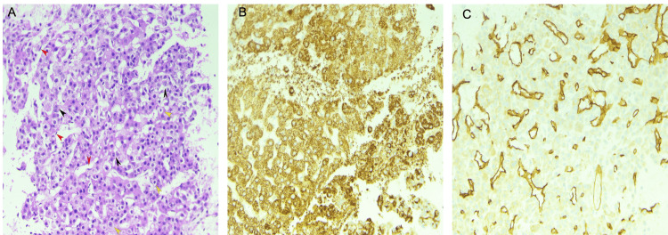
Figure 2. (A) FNA cell block section shows well to moderately differentiated HCC arranged in cords of neoplastic cells, few cells with large pleomorphic nuclei and nucleoli (black arrow) interspersed with clear cells (yellow arrow) and flat endothelial cells wrapping the trabeculae (red arrow) (H&E 10x). (B) Hep Par 1 immunostain positive in HCC tumor cells (IHC 10x). (C) CD34 immunostain highlighting the endothelial cell proliferation in HCC (IHC 10x).
FNA: Fine-needle aspiration; HCC: Hepatocellular carcinoma.

