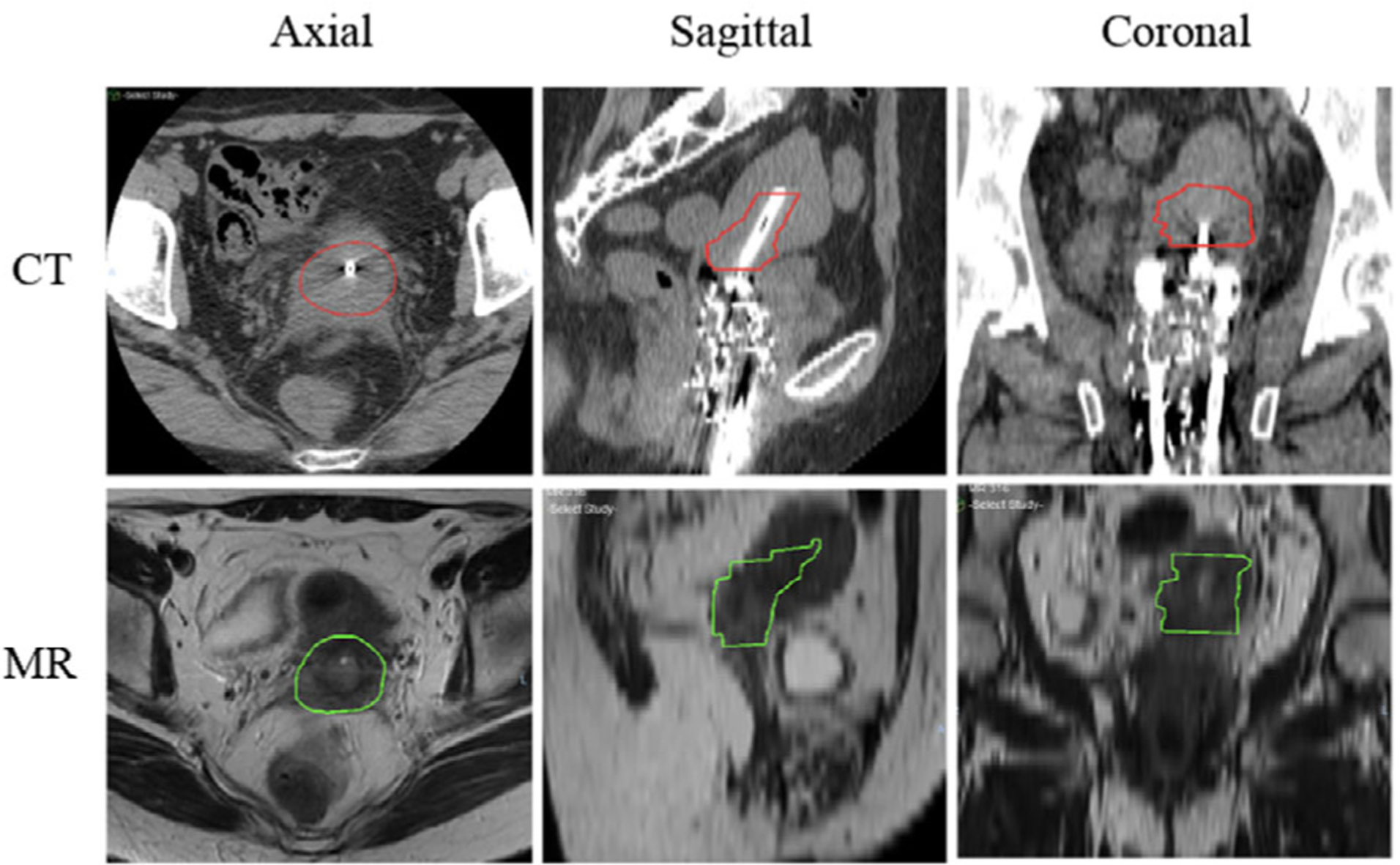
FIGURE 1.

Axial, sagittal, and coronal views of computed tomography (CT) and MR for a representative patient, which show the comparison of location and size of preimplant MR-based high-risk clinical target volume (HR-CTVMR) and postimplant planning-CT-based HR-CTVCT
