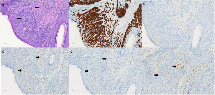
FIGURE 3.
(a) In epidermis and dermis, there are abundant pale-staining cytoplasmic, which are flaky, small nests or scattered cells in HE staining, involving both the dermis and skin appendages. Left arrow: Paget cell. Right arrow: infiltrating adenocarcinoma. (b) Positive expression of CK (7) in EMPD section 100×. (c) Negative expression of CK (20) in EMPD section 100×. (d) Negative expression of CD (56) in EMPD section 100×. (e) Negative expression of ChrA in EMPD section 100×. (f) Focal positive expression of synaptophysin in EMPD section 100×.

