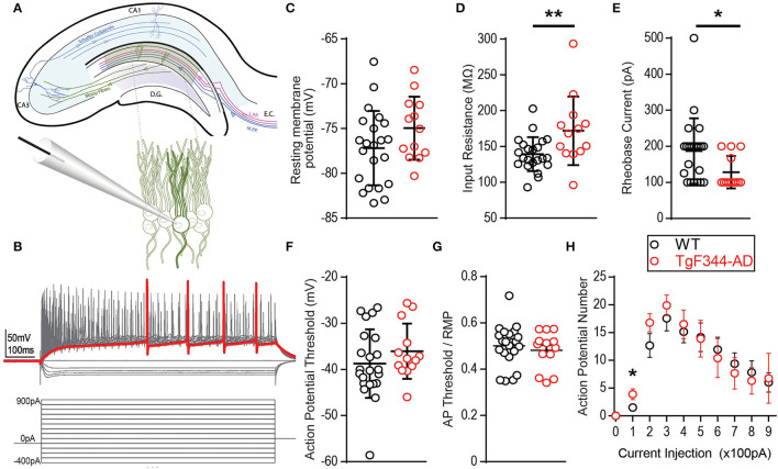
Figure 1.
Membrane properties are altered in TgF344-AD dentate granule cells (DGCs). (A) Schematic of the trisynaptic circuit in a coronal slice from rat hippocampus (top) with expanded recording setup (bottom). (B) Illustration of the experimental protocol: 14 serial 100 pA hyperpolarizing to depolarizing current steps at 800 ms duration from −400 pA to +900 pA. The red trace represents the first depolarizing current step to fire and action potential. (C) Mean resting membrane potential is not different between wild type (Wt) (black, 22 cells/8 animals) and TgF344-AD female rats (red, 13 cells/6 animals; p > 0.05, but a higher fraction in Wt has values below −80 mV). (D) Input resistance is significantly decreased in TgF344-AD DGCs (Wt n = 22 cells/8 animals; Tg, n = 13 cells/6 animals; **p ≤ 0.01). Data represent mean ± SEM. Significance is determined using the unpaired t-test. (E) The first depolarizing current step to elicit and action potential, Rheobase (pA), is significantly decreased in TgF344 compared with Wt (*p < 0.05). (F) Action potential threshold is not different between genotypes (p > 0.05). (G) When AP threshold is normalized to RMP, there remains no difference between genotype (p > 0.05). (H) AP number was enhanced for TgF344-AD rat DGCs at 100 pA current injection (p < 0.05) but not at higher current injections. For all panels, Wt, n = 22 cells/8 animals; Tg, n = 13 cells/6 animals). Data represent mean ± SEM. Significance is determined using the unpaired Student's t-test.

