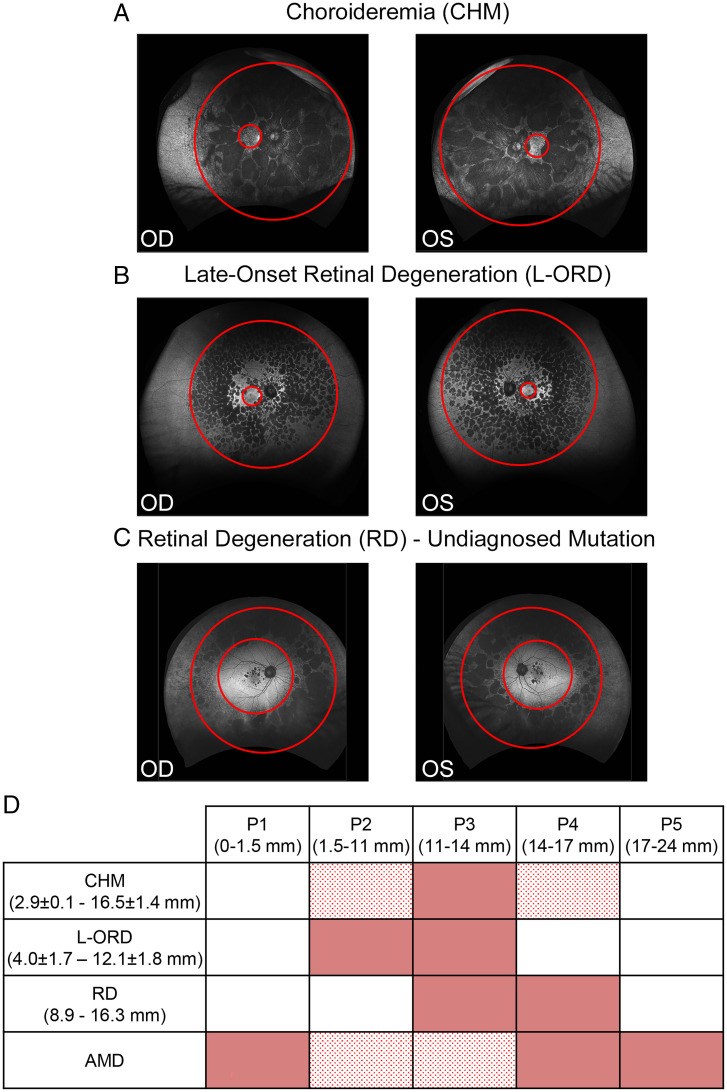
Fig. 6.
Different retinal diseases affect different RPE subpopulations. (A–C) Right (OD) and Left (OS) eye fundus images of patients affected by CHM (A) and L-ORD (B) and a patient with RD with an undiagnosed mutation (C) with RPE degeneration in different regions of the eye. Red circles higlight the inner and outer boundaries of the areas of retinal degeneration. (D) Table summarizes defects in RPE subpopulations in different forms of RDs [mean ± SD of radii (in millimeters) of inner and outer limits of atrophic regions]. Red boxes correspond to fully affected RPE subpopulations, dotted red boxes correspond to partially affected subpopulations, while white boxes indicate unaffected subpopulations.

