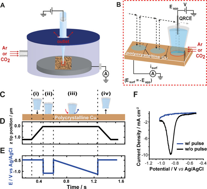
Figure 1.
Schematics of the SECCM experimental setup and scanning protocol (not to scale). (A) Nanopipette probe, filled with the 10 mM KHCO3 electrolyte and containing a QRCE, in the environmental cell hosting the substrate (polycrystalline Cu embedded in a block of carbon, as schematized through the displayed grain boundaries). Flow of humidified gas (Ar or CO2) through the cell, saturating the nanodroplet meniscus and the solution in the lower part of the nanopipette. (B) Expanded view of the nanopipette tip region, illustrating the hopping-mode protocol used in this work. The trajectory of the tip during the scan is shown by the dotted lines with arrowheads, and the area wetted by the nanodroplet (working electrode area) is shown as blue circles. Linear sweep voltammetry (LSV) measurements were made at each hop position by sweeping the potential, Eapp, of the QRCE and measuring the current, isurf, at the working electrode. Substrate potential, Esurf = −Eapp. (C) Stepwise events (i–iv) at each hop of the scan, with the corresponding plot of (D) z-displacement of the pipet meniscus from the surface (meniscus contact defined as z = 0 μm) and (E) synchronous potential waveform. In (C–E), (i) the tip approaches the surface with Esurf = −0.5 V upon meniscus contact; (ii) Esurf is stepped to −1.05 V for 150 ms to reduce the native surface layer on the Cu substrate; (iii) Esurf then stepped back to −0.5 V and linearly swept to −1.05 V (1 V/s); and (iv) Esurf stepped to −0.5 V, and the tip retracted away from the surface. (F) Comparison of linear sweep voltammograms from the protocol described in C–E (blue trace) to analogous LSV without the electrochemical pre-treatment step (black trace).

