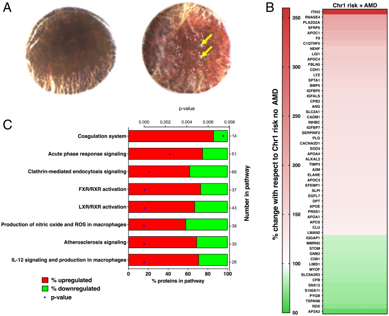
Fig. 1.
(A) Representative submacular tissue punches after removal of neurosensory retina from donors with no AMD (Left) and early AMD (Right); yellow arrows indicate drusen that had a diameter of greater than 63 µm. (B) Proteins with significantly changed levels when comparing submacular stromal tissue punches from Chr1 high-risk donors with and without AMD changes; red indicates increased with AMD, and green indicates decreased with AMD. (C) Ingenuity pathways analysis showing altered canonical pathways in submacular stromal tissue punches from Chr1 risk donors comparing with and without AMD. FXR, farnesoid X receptor; LXR, liver X receptor; ROS, reactive oxygen species.

