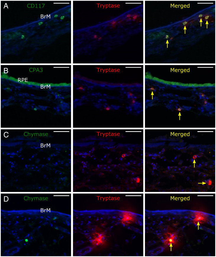
Fig. 2.
Representative confocal micrograph z-stack projections of immunolabeled submacular sections labeled for (A) CD117 (green) and tryptase (red), (B) CPA3 (green; in this section, the RPE is present, which is autofluorescent, especially in the green channel) and tryptase (red), and (C and D) chymase (green) and tryptase (red) in C, showing MCT-type mast cells and in D, showing the halo of tryptase labeling surrounding degranulating MCTC-type mast cells. Sections were counterstained with Hoescht nuclear stain (blue), which stained Bruch’s membrane (BrM) and stroma of the inner choroid as well as cell nuclei. Yellow arrows point to mast cells. (Scale bars: 50 µM.)

