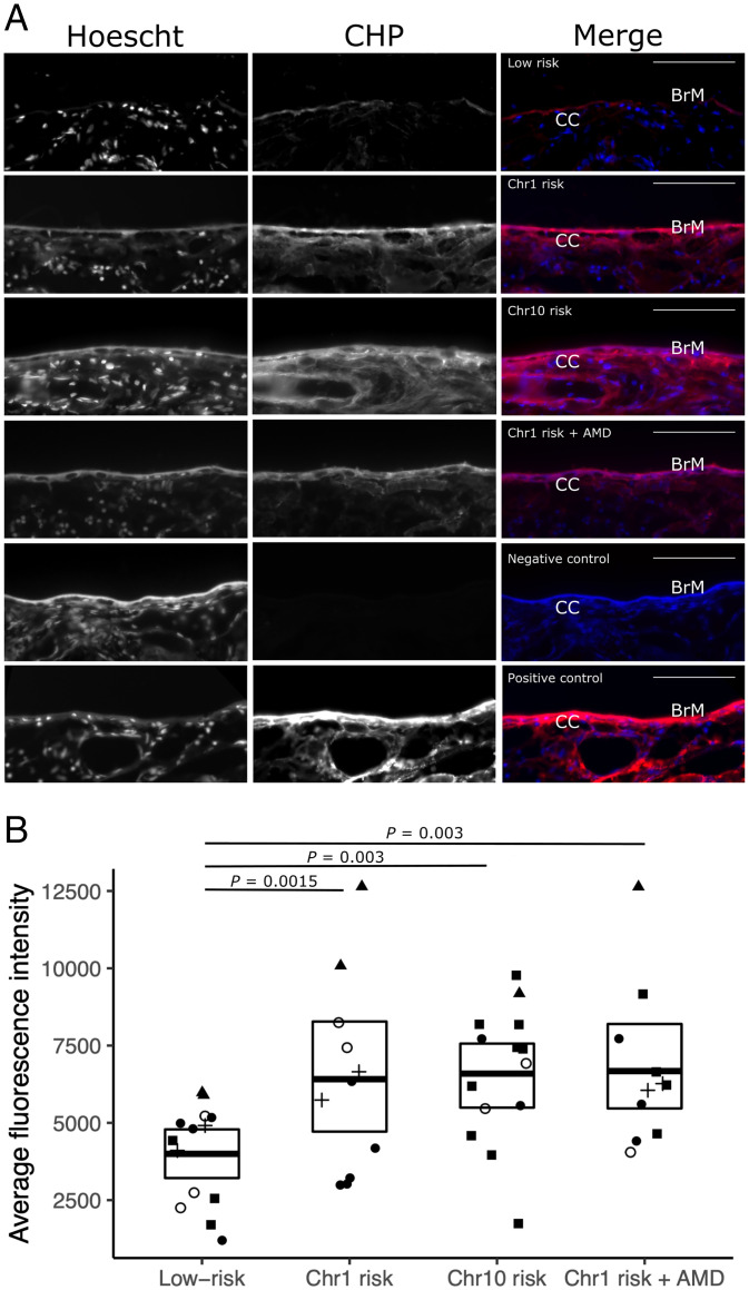
Fig. 4.
(A) Submacular sections labeled with denatured CHP linked to Cy5 (CHP) and Hoescht nuclear stain (which labeled the Bruch’s membrane [BrM] and the choroidal stroma as well as cell nuclei) showing representative sections of genetically low risk, Chr1 risk, Chr10 risk, and Chr1 risk with AMD along with negative and positive controls. In merged images, CHP is red, and Hoescht nuclear stain blue; BrM and the choriocapillaris (CC) are labeled. (Scale bars: 50 µm.) (B) Quantification of denatured CHP binding to BrM, where the fold change of fluorescence intensity within a 5 µM width line over BrM was analyzed comparing low risk with Chr1 risk, Chr10 risk, and Chr1 risk with AMD. Significantly different P values are shown, where P < 0.05 as determined using a Poisson generalized linear mixed effects models; different experiments are indicated by different symbols.

