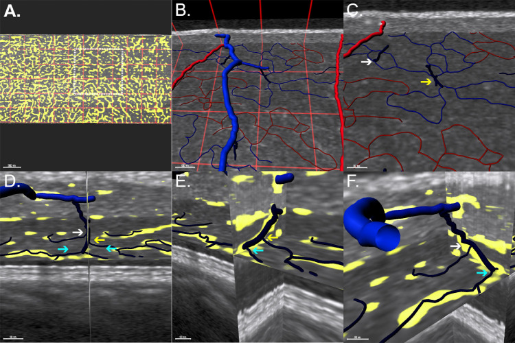
Figure 3.
Morphologic characteristics of deep vascular complex outflow pathways. (A) En face view of the ICP. The image is divided by a red grid comprising squares sized 330 × 330 µm. A white dashed square highlights the region shown in B and C. (B) Superior view includes superficial larger vessels with ICP and ICP draining venules. (C) Magnified view showing two distinct configurations of capillary convergence. The white arrow indicates the convergence of two capillaries draining a small region, and the yellow arrow indicates a draining venule resulting from the convergence of four capillaries. (D) Coronal view of a venule originating from a DCP vortex. A plane spanning the XY axis is centered on the venule. (E, F) Orthogonal views demonstrate that DCP capillaries join the draining venule at different levels on the XY plane (different-colored arrows), resembling a spiral staircase morphology.

