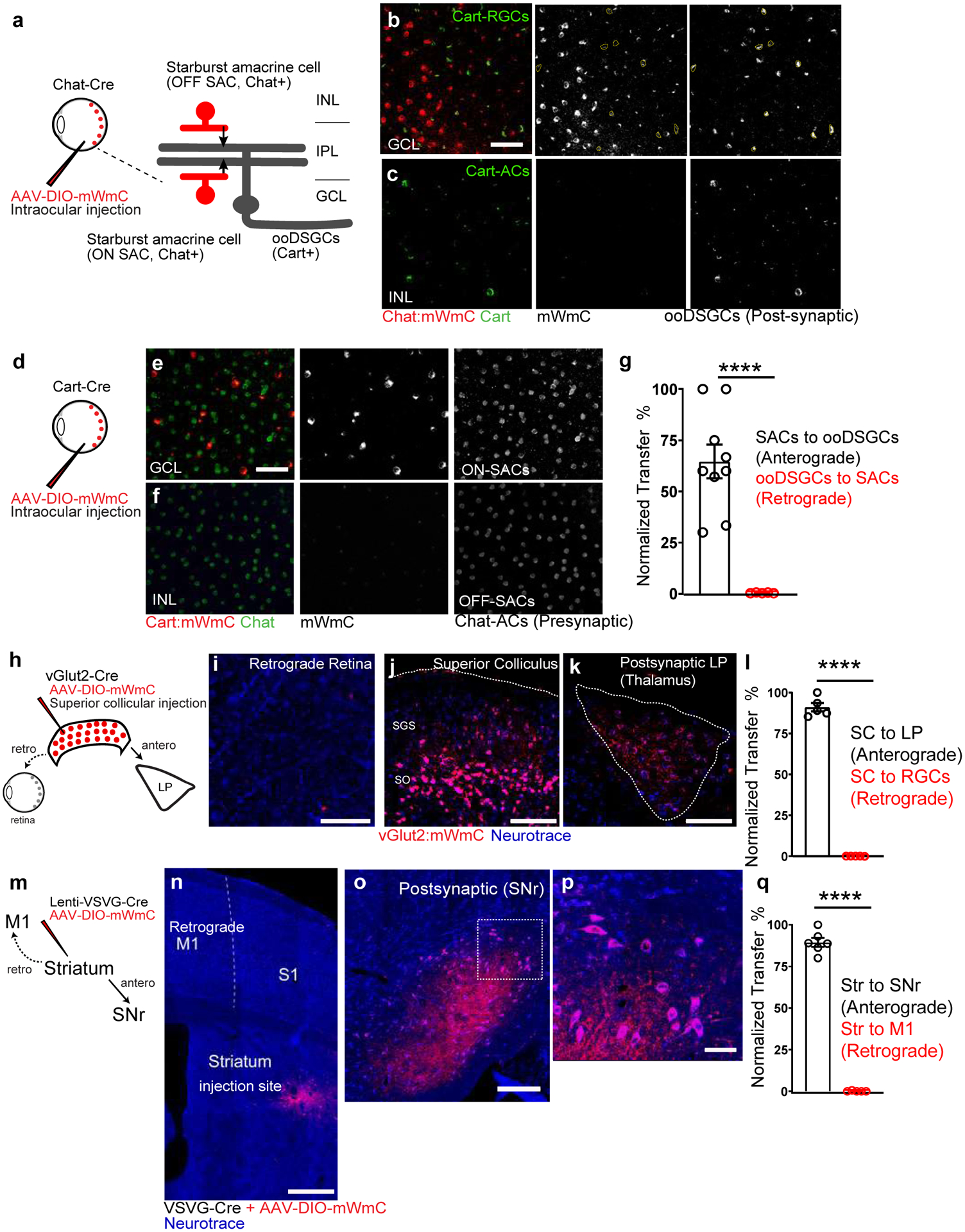
Fig. 3. Anterograde but not retrograde transfer of mWmC in retinal and brain circuits.

a-c, (a) Intraocular injections of mWmC into Chat-Cre led to (b) expression of mWmC within Starburst Amacrine Cells (SACs, red) within the GCL as starter cells. ooDSGCs (Cart-positive, green), which receive direct input from SACs, were labeled by mWmC through anterograde transfer (mWmC double-positive cells labeled with yellow-dotted circles). No signal was detected in the INL, indicating that bipolar cells were not labeled from SACs retrogradely (c), 8 times each experiment was repeated independently with similar results. d-f, (d) Intraocular injections of mWmC into Cart-Cre led to (e) Efficient transduction of ooDSGCs as starter cells for anterograde tracing into the brain (Fig. 5). No retrograde spread was seen into SACs (Chat-positive) or bipolar cells in either the GCL or INL (f), 5 times each experiment was repeated independently with similar results. Scale bars: b, c, e, f, 50μm. g, Quantifications of the anterograde transfer ratio from SACs to ooDSGCs (black) n=9 biologically independent samples, versus retrograde transfer ratio from ooDSGCs to SACs (red), n=5 biologically independent samples. ****, p<0.0001, two-sided Student’s t-test. Within each retina region, 43±6% of the Cart-positive RGCs received mWmC transfer from SACs. h-l, (h) Introduction of mWmC into excitatory SC neurons (vGlut2-positive) for anterograde and retrograde transfer tests. (j) Efficient start neurons at the SC demonstrated very few retrograde spread back to the retina (i), but specific and efficient anterograde transfer onto the LP of the thalamus (k). Scale bars: (i, 50μm, j, 200μm, k, 100μm). l. Quantifications of the anterograde transfer ratio from SC to LP (black) versus retrograde transfer ratio from SC to RGCs (red), n=5 animals. ****, p<0.0001, two-sided Student’s t-test. Within each SC starting region, 81±6% of the NeuN-positive SC neurons were infected with mWmC; within the LP recipient neuron regions, 86±4% received mWmC transfer from the SC. m-q, (m) Introduction of Cre-dependent mWmC with VSVG-lentivirus expressing Cre into the dorsal lateral striatal neurons for anterograde and retrograde transfer tests. (n) Efficient start neurons at the striatum demonstrated no retrograde transfer back to M1 on the same brain slice. In contrast, mWmC displayed specific anterograde transfer onto the Substantia Nigra (o, SNr) as zoomed-in (p). Notably, axons are also filled by mWmC, in addition to somata filling. Scale bars: (n, 500μm, o, 100μm, p, 50μm). q. Quantifications of the anterograde transfer ratio from the striatum to SNr (black) versus retrograde transfer ratio from the Striatum to M1 (red), mWmC-positive neurons, n=6 animals ****, p<0.0001, two-sided Student’s t-test. Within the SNr recipient neuron regions, 89±2% received mWmC transfer from the Striatum. All data in this figure are presented as mean ± SEM.
