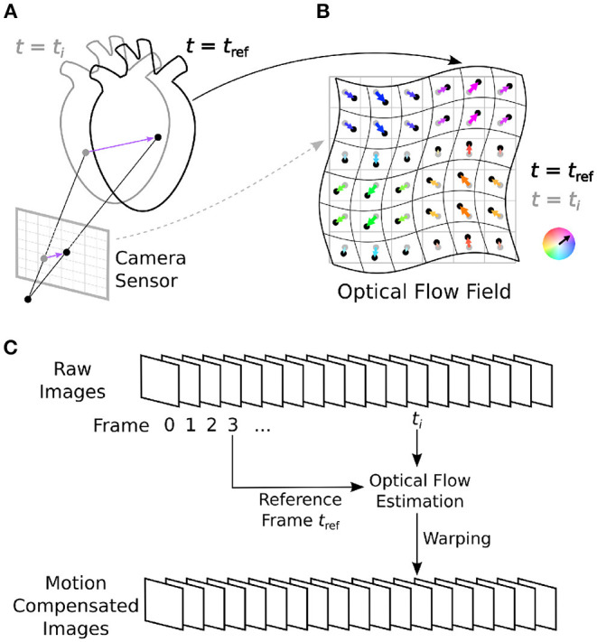
Figure 1.

Optical mapping combined with numerical motion tracking for measurement of an action potential or calcium waves in moving and deforming cardiac tissues, such as an isolated heart during voltage-sensitive optical mapping. (A) Two-dimensional in-plane displacements on the camera sensor depict the motion of the tissue between two video frames from time ti to time tref. (B) Optical flow field describing the deformation of the imaged tissue pixel-by-pixel. In Figure 10 and Supplementary Figures S1, S2, the displacement vector fields are HSV color-coded: the orientation and magnitude of the displacements are depicted by hue (color) and saturation, respectively. (C) Numerical motion tracking in a sequence of video images with a calculation of the displacements with respect to a reference frame (first frame or any arbitrary frame).
