Figure 4.
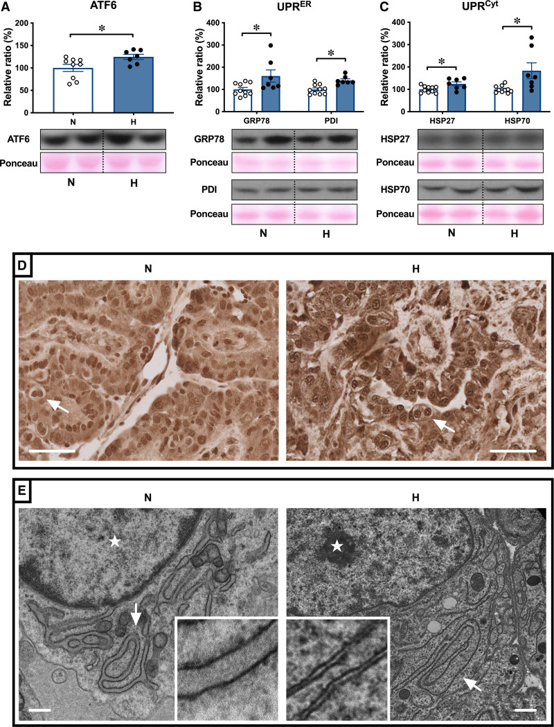
Hypoxic pregnancy activates the placental UPR (unfolded protein response). Values are mean±SEM for the relative ratio of the placental levels of ATF6 (activating transcription factor 6; A), GRP78 (glucose-related protein 78) and PDI (protein disulfide isomerase; B), and HSP27 (heat shock protein 27) and HSP70 (heat shock protein 70; C). Groups are normoxic (N; ○, n=9–10) and hypoxic (H; ●, n=7). Significant differences (P<0.05) are *N vs H, Student t test for unpaired data. In the placenta, ATF6 localizes to the nuclei (D), with more prominent nuclear staining in H compared with N placentae. Pictured (D) trophoblast containing binucleate cells (arrows); scale bar=50 µm. Change in trophoblast endoplasmic reticulum (ER) structure was examined by transmission electron microscopy (E). Representative images taken at ×5000 magnification are shown. Arrows indicate the location of ER and stars indicate the location of the nucleus; scale bar=500 nm.

