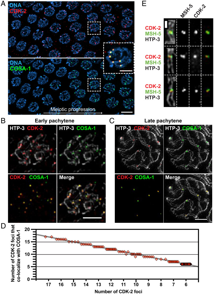
Fig. 1.
CDK-2 colocalizes with COSA-1 both at early recombination intermediates and at late pachytene crossovers. (A) Immunofluorescence images of a whole-mount gonad from a worm strain that expresses CDK-2::AID::3×Flag and GFP::COSA-1. (Scale bar, 5 μm.) Inset, CDK-2 and COSA-1 colocalizing in six bright foci in nuclei following transition to late pachytene. (Scale bar, 2 μm.) (B and C) Full projections of SIM images of spread gonads showing the staining for HTP-3 (white), CDK-2 (red), and COSA-1 (green) from early pachytene (B) and late pachytene nuclei (C). (Scale bars, 2 μm.) (D) Quantification of CDK-2 foci in pachytene nuclei and their colocalization with COSA-1. Each diamond represents a nucleus; the gray line indicates perfect colocalization. (E) Representative SIM images of individual crossover–designated sites showing CDK-2 singlet foci localizing together with MSH-5 doublets. (Scale bar, 500 nm.)

