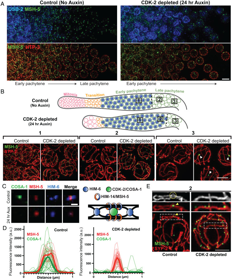
Fig. 3.
CDK-2 is required for stabilizing crossover–specific recombination intermediates. (A) Animals expressing CDK-2::AID::3×Flag and TIR1::mRuby were treated with or without 1 mM auxin for 24 h after L4. Dissected gonads were spread and stained for DSB-2 (blue), MSH-5 (green), and HTP-3 (red). (Scale bar, 5 μm.) (B) Top, diagram illustrating the effect of CDK-2 depletion on meiotic progression. The DSB-2–positive nuclei (shown in blue) represent nuclei in early pachytene. Bottom, representative SIM images of nuclei from the indicated regions (1, 2, or 3) of spread gonads from control and CDK-2–depleted worms as indicated in the diagram above. White arrowheads in 3 indicate large MSH-5 aggregates. (Scale bar, 3 μm.) (C) Representative fluorescent images of recombination sites in late pachytene from control versus CDK-2–depleted germline. Stainings for MSH-5, HIM-6, and COSA-1 are shown. (Scale bar, 400 nm.) A schematic depicting the hypothesized architecture of recombination factors at the crossover–designated site is shown on the right. (D) Line scan profiles of MSH-5 (red) and COSA-1 signals (green) at recombination sites in late pachytene nuclei from control (n = 26) and CDK-2–depleted germlines (n = 26). Thin lines are individual traces, and thick lines are averages; a.u., arbitrary units. (E) Representative SIM images of spread gonads from region 2 in the diagram above and a segment of SC stretch from control and CDK-2–depleted animals. MSH-5 (green) and SYP-2 (red) stainings are shown. The yellow circle and arrowhead in the control indicate the SC bubble at the crossover site. (Scale bar, 2 μm.)

