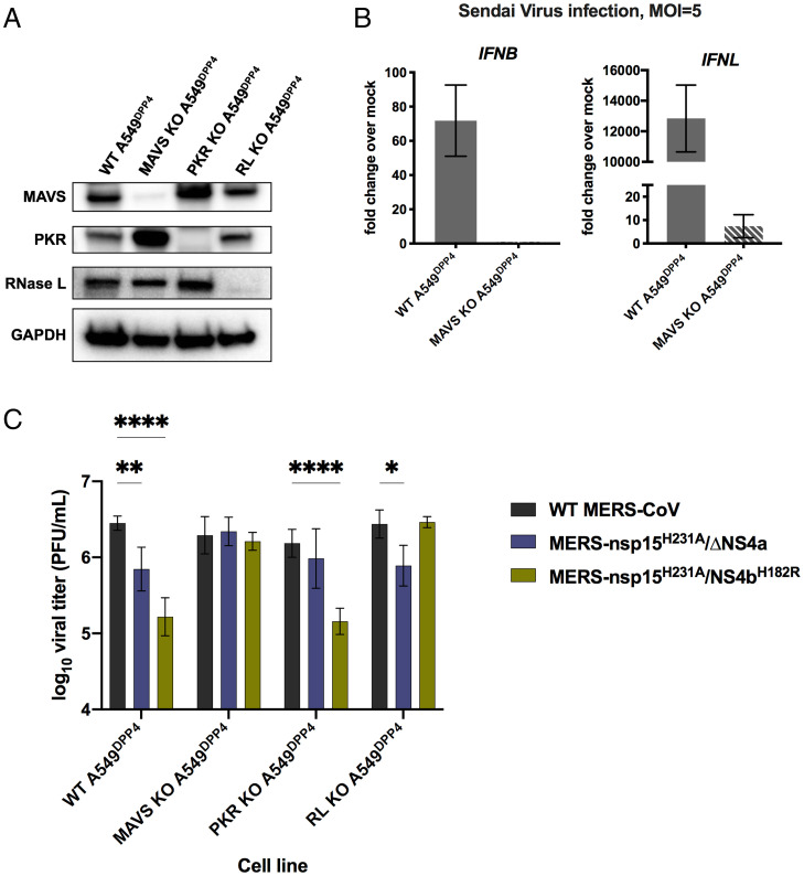
Fig. 5.
KO of innate immune pathways rescues attenuation in MERS-nsp15H231A/ΔNS4a and MERS-nsp15H231A/NS4bH182R. (A) Cell lysates of WT A549DPP4 as well as MAVS KO, PKR KO, and RNase L KO A549DPP4 were collected, and proteins were separated by SDS/PAGE and immunoblotted with antibodies against MAVS, PKR, RNase L, and GADPH. (B) WT A549DPP4 and MAVS KO A549DPP4 cells were infected with Sendai virus at MOI = 5, and total cellular RNA was harvested at 18 hpi. Expression of IFNL1 and IFNB was quantified by qRT-PCR, and CT values were normalized to beta-actin and shown as fold change over mock using the formula 2-Δ(ΔCT). Data are displayed as means ± SD. (C) A549DPP4 cell lines: WT, MAVS KO, PKR KO, and RNase L KO were infected in triplicate at MOI = 1, and supernatant samples were collected at 48 hpi. Viral replication was quantified by plaque assay. Data are displayed as means ± SD. Statistical significance of differences in viral replication for each recombinant virus compared to WT MERS-CoV was calculated by repeated measures two-way ANOVA: *P ≤ 0.05; **P ≤ 0.01; ****P ≤ 0.0001. Data that were not statistically significant are not labeled. Data are from one representative of three independent experiments.

