Fig. 2.
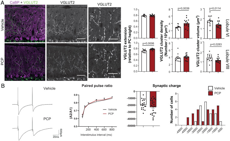
Neonatal PCP administration leads to long-lasting changes in CF/PC connectivity. (A) CF presynaptic boutons were immunostained with an anti-VGLUT2 antibody (green) and PCs and their dendritic tree were stained with an anti-calbindin antibody (magenta) in parasagittal cerebellar sections from P30 vehicle- and PCP-treated mice. High-magnification images of VGLUT2 clusters are shown for both conditions. Quantifications of the extension of the CF synaptic territory, mean volume of the VGLUT2 puncta, and their mean density were performed in lobules VI and VIII (mean ± SEM is represented, with P values when they are significant; unpaired Student’s t test for the volume, Welch’s t test for the extension and density; vehicle: n = 13 to 14 animals; PCP: n = 15 to 16 animals). (Scale bars: 50 µm; high-magnification images, 20 μm.) (B) PC responses after CF stimulation were recorded using patch clamp. Representative traces of responses obtained in PCs from lobule VI after paired-pulse stimulation (50-ms interval) are shown for both conditions. No difference in the paired-pulse ratio (ratio between the amplitude of the second response and the first response A2/A1) was detected regardless of the stimuli interval between PCP mice and vehicle mice (Kolmogorov–Smirnov test). The mean synaptic charge after CF stimulation is not significantly different between the PCP and vehicle conditions (vehicle: n = 19 cells/9 mice and PCP: n = 23 cells/9 mice; unpaired Student’s t test), but a change in the distribution is visible with a shift toward higher charges in the PCP condition.

