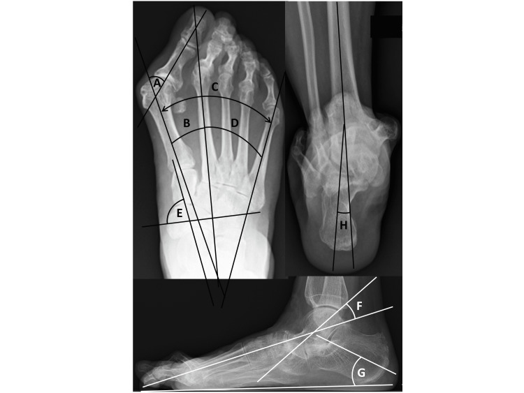
Figure 1. Radiography to measure parameters of foot deformity. Dorsoplantar, anteroposterior weight-bearing radiographs.
A) Hallux valgus (HV) angle.
B) Intermetatarsal angles between first and second metatarsals (M1-M2A).
C) Intermetatarsal angles between first and fifth metatarsals (M1-M5A).
D) Intermetatarsal angles between second and fifth metatarsals (M2-M5A).
E) Pronated foot index (PFI), measured as the angle between the short axis of the navicular and the long axis of the talus (normal, >65°). Lateral weightbearing radiographs.
F) Talo-first metatarsal angle (Meary angle).
G): Calcaneal pitch angle. Radiographs were taken in a weight-bearing position. Covey method view.
H) Tibio-calcaneal angle (TCA).

