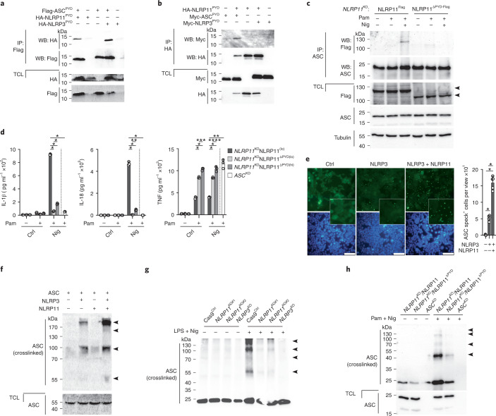
Fig. 4. NLRP11PYD recruits ASC and is necessary for efficient ASC polymerization.
a,b, Immunoprecipitation with immobilized anti-Flag (a) and anti-HA (b) antibodies, and TCLs were analyzed by immunoblot for HA-, Myc- and Flag-tagged proteins after transient transfection of HEK293 cells with Flag-ASCPYD, HA-NLRP11PYD and HA-NLRP3PYD (a) and HA-NLRP11PYD, Myc-ASCPYD and Myc-NLRP3PYD (b), as indicated. c, Immunoprecipitation with immobilized anti-ASC antibodies from TCL of NLRP11KO cells restored with NLRP11Flag or NLRP11ΔPYD-Flag left untreated, primed with Pam3CSK4 (1 μg ml−1, 2 h) and primed and activated with nigericin (5 μM, 10 min); immunoprecipitates and TCLs were analyzed by immunoblot for Flag, ASC and tubulin loading control. Arrowheads indicate the correct-size protein. d, IL-1β, IL-18 and TNF ELISA from SNs of NLRP11KO cells restored with low-expressing NLRP11Flag cells (NLRP11lo, NLRP11ΔPYD(lo)), high-expressing NLRP11ΔPYD(hi) and ASCKD cells left untreated, primed with Pam3CSK4 (1 μg ml−1, 4 h) and primed and activated with nigericin (5 μM, 25 min) (n = 3, mean ± s.d.); *P < 0.0001, **P = 0.0002, ***P = 0.0007, ****P = 0.0038. The dotted line indicates that for ASCKD, only the Pam3CSK4 + nigericin group is shown. e, Fluorescence microscopy of EGFP and DAPI in HEK293ASC-EGFP cells transiently transfected with empty vector (Ctrl), NLRP3 and NLRP11 as indicated (left) and quantification of ASC speck+ cells per view (right). (n = 5, mean ± s.d.); *P < 0.0001, scale bars, 100 μm. f, immunoblot for ASC of crosslinked TCL from above cells. Arrowheads indicate oligomers. g,h, Immunoblot for ASC of TCLs and crosslinked TCLs from Cas9Ctrl, NLRP11KO#1, NLRP11KO#2 and NLRP3KO cells left untreated or primed with LPS (200 ng ml−1, 4 h) and activated with nigericin (5 μM, 20 min) (g) and NLRP11Flag, NLRP11ΔPYD-Flag and ASCKD cells left untreated or primed with Pam3CSK4 (1 μg ml−1, 4 h) and activated with nigericin (5 μM, 15 min) (h). Arrowheads indicate oligomers.

