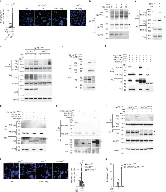
Fig. 6. NLRP11 acts as a scaffold for NLRP3 inflammasome assembly.
a, Confocal microscopy of PLA (green) between NLRP3 and Flag and DAPI using PMA-differentiated CtrlFlag and NLRP11Flag cells left untreated, primed with LPS (200 ng ml−1, 4 h) and primed and activated with nigericin (5 μM, 20 min); scale bar, 50 μm; (right), and quantification of PLA+ cells per view (left) (n = 4, mean ± s.d.); *P = 0.0004; **P = 0.0001. b, Immunoprecipitation with immobilized anti-NLRP3 antibodies using TCLs from untreated, LPS-primed (200 ng ml−1, 4 h) and primed + nigericin (5 μM, 20 min and 45 min) activated THP-1 cells and immunoblot of immunoprecipitates and TCLs for NLRP11 and NLRP3. Arrowheads indicate the correct-size proteins. c, Immunoprecipitation with immobilized anti-Flag antibodies using SNs of NLRP11Flag cells primed with LPS (200 ng ml−1, 4 h) and primed and activated with nigericin (5 μM, 10 min) and immunoblot of immunoprecipitates and TCLs for NLRP3 and Flag. d, Immunoprecipitation with immobilized anti-NLRP3 antibodies using TCLs from NLRP11KO and ASCKD THP-1 cells restored with NLRP11-Flag left untreated, LPS-primed (200 ng ml−1, 2 h) and primed and activated with nigericin (5 μM, 10 min), and immunoblot of immunoprecipitates and TCLs by immunoblot for Flag, NLRP3, ASC and tubulin loading control. e–h, Immunoprecipitation with immobilized anti-HA (e), anti-Flag (f,g) and anti-EGFP (h) antibodies using TCLs from HEK293 cells transiently transfected with Flag-NLRP11NACHT and HA-NLRP3NACHT (e) ; Flag-NLRP11NACHT, HA-NLRP3NACHT, HA-NLRP12NACHT and HA-NOD1NACHT (f); Flag-NLRP11NACHT, Myc-NLRP3, Myc-NLRC4 and Myc-pyrin (g); and EGFP-NLRP11LRR, Myc-NLRP3, Myc-NLRC4 and Myc-pyrin as indicated (h); and immunoblot of immunoprecipitates and TCLs for HA-, Flag-, Myc- and EGFP. Asterisk denotes modified pyrin (g,h). The gap in the TCLs in panel h marks an empty lane between NLRC4 and pyrin. i, Immunoprecipitation with immobilized anti-NLRP3 antibodies using TCLs from NLRP11Flag and NLRP11ΔPYD- Flag cells left untreated, primed with Pam3CSK4 (1 μg ml−1, 2 h) and primed and activated with nigericin (5 μM, 10 min) and immunoblot of immunoprecipitates and TCLs for Flag, NLRP3 and tubulin loading control. j, Confocal microscopy of PLA (red) between NLRP3 and caspase-1 and DAPI of PMA-differentiated Cas9Ctrl, ASCKD and NLRP11KO cells primed with LPS (200 ng ml−1, 4 h) and primed and activated with nigericin (5 μM, 20 min); scale bar, 50 μm; (left), and quantification of PLA+ cells per view (right) (n = 4, mean ± s.d.); *P = 0.0021; **P = 0.0006, ***P = 0.0003. k, IL-1β ELISA of SNs from Cas9Ctrl and NLRP3KO cells stably expressing NLRP11-Flag left untreated, primed with LPS (200 ng ml−1, 4 h) and primed and activated with nigericin (5 μM, 30 min) (n = 3, mean ± s.d.); *P = 0.0006, **P = 0.0003.

