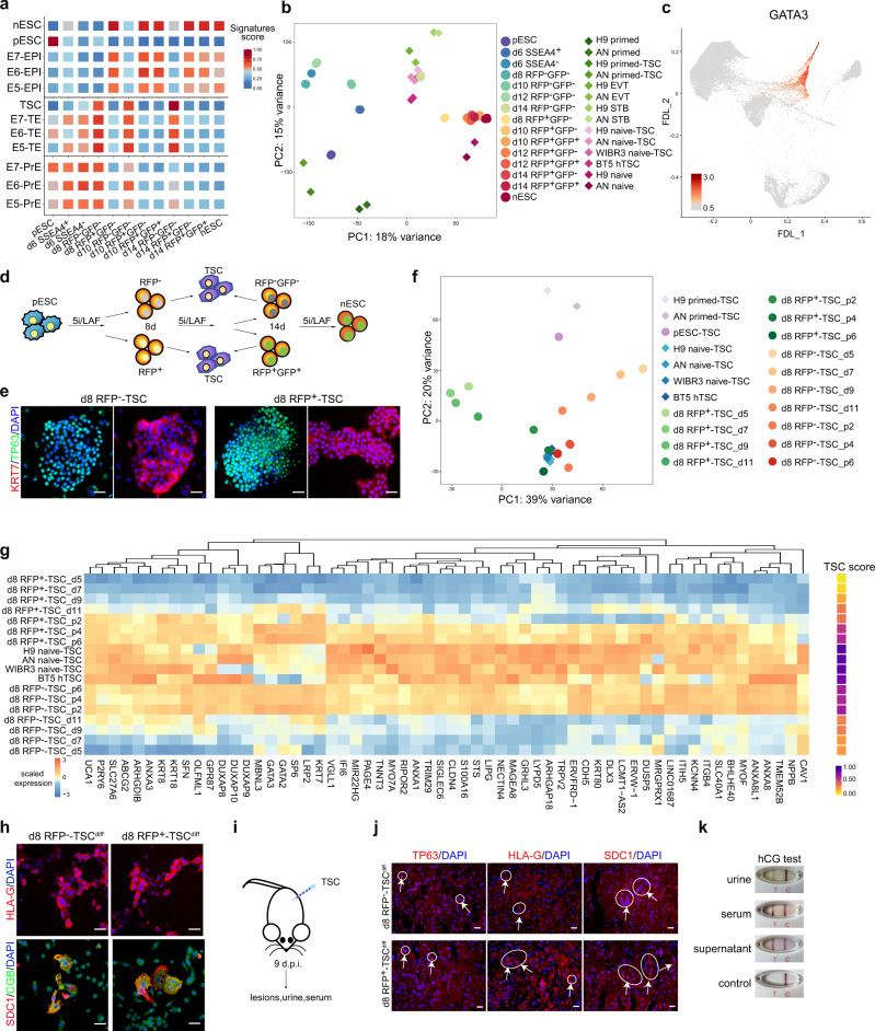
Fig. 4. TE signatures during the primed-to-naive transition.
a EPI, TE, PrE and trophoblast stem cell (TSC) signature scores of the primed-to-naive transitioning intermediates. b PCA of the bulk RNA-seq datasets (circles) from the primed-to-naive transitioning intermediates with published RNA-seq (diamonds) datasets32. n ≥ 2. c Expression of GATA3 in FDL. d Experimental design for the induction of TSCs from the primed-to-naive intermediates. e TP63 and KRT7 immunostaining of TSCs derived from day 8 RFP− and day 8 RFP+ cells during the primed-to-naive transition. Scale bars, 20 μm. Representative images from n = 3. f PCA of the bulk RNA-seq datasets (circles) from the transitioning intermediates-derived TSCs with published RNA-seq (diamonds) datasets32. n ≥ 2. g Heatmap (left) and TSC score (right) showing the expression levels of representative TSC-related genes during the TSC derivation process from RFP− and RFP+ transitioning intermediates on day 8. Source data are provided as a Source Data file. h HLA-G, SDC1 and CGB immunostaining of extravillous trophoblast (EVT) (upper) and syncytiotrophoblast (ST) (lower) cells, respectively. EVT and ST cells were differentiated from day 8-RFP− and day 8-RFP+ cell-derived TSCs. Scale bar, 20 μm. Representative images from n = 3. i Representation of day 8-RFP− and day 8 RFP+ cell-derived TSC engraftment assay by injection into NOD-SCID mice. j Immunostaining of TP63, HLA-G and SDC1 in the lesions collected from day 8-RFP− and day 8 RFP+ cell-derived TSC engrafts in NOD-SCID mice. No lesions were evident in the vehicle controls. Scale bar, 20 μm. Representative images from n = 3. k Representative positive results for the hCG pregnancy test performed on urine samples, serum samples, and ST cell culture supernatant collected from day 8-RFP− cell -derived TSCs. Source data are provided as a Source Data file.

