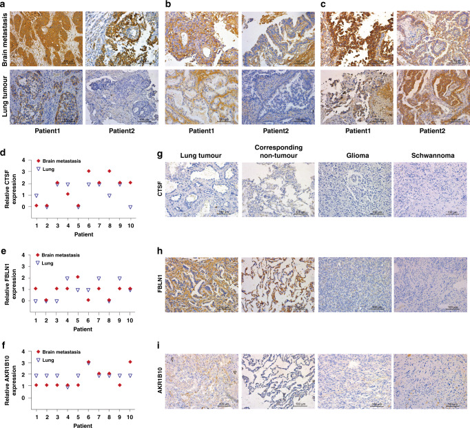
Fig. 3. Tissue expressions of CTSF, FBLN1 and AKR1B10 in cohort 3.
Cathepsin F (CTSF) (a), Fibulin-1 (FBLN1) (b) and Aldo-keto reductase family 1 member B10 (AKR1B10) (c) representative immunohistochemistry (IHC) staining of paired primary lung tumour and brain metastasis (BM) tissues from 2 patients in cohort 3. A representative area is selected. Relative expressions of CTSF (d), FBLN1 (e) and AKR1B10 (f) in primary lung tumour and BM tissues of 10 patients are shown. 0: negative, 1: weak positive, 2: positive, 3: strong positive. Representative IHC staining for CTSF (g), FBLN1 (h) and AKR1B10 (i) of patients from each control in cohort 3. Corresponding non-tumour is matched with non-small cell lung cancer patient without BM.

